13125
0
In this article:
- Causes
- Age characteristics
- What pathologies can a large fontanelle indicate?
- When it's dangerous
- How does a large fontanel close?
- Rules for caring for a large fontanel
- Useful video about fontanel in a newborn
Every newborn child has at least one fontanelle on its skull, which is represented by a non-ossified area of membranous tissue. Performing a huge number of functions, these structures, by increasing or decreasing, can reflect some congenital and acquired pathologies in infants. Therefore, a large fontanelle in a newborn requires dynamic monitoring from both the parents and the pediatrician.

During each monthly examination, the local pediatrician should assess the size and condition of the existing fontanels, which will allow him to suspect a developing disease in time. However, we should not forget that an increase in the anterior fontanel in some cases is within the normal range.
Causes
The anterior fontanel itself is large in size compared to the others and is characterized by gradual closure. Only after measuring it with a ruler and comparing the child’s age with the standard can one judge whether the fontanelle is increasing or decreasing.
Some physiological reasons affecting the size of the cartilaginous portion of the child’s skull:
- Transient conditions that occur in the mother during pregnancy. Hypovitaminosis and calcium deficiency due to poor nutrition, mild anemia - all this can contribute to the enlargement of the anterior fontanel;
- Genetic predisposition. If the child is calm, eats well, gains adequate weight, but, like many of his relatives, has a large fontanelle, then this is included in the concept of the norm and is considered a family feature;
- Baby's diet and feeding patterns. As you know, breast milk is an ideal balanced product that allows an infant to fully develop and grow. Even the most adapted and expensive artificial formula can never compare with breast milk. Therefore, in children who are on mixed or exclusively artificial nutrition, large fontanel sizes are often detected;
- Immaturity and prematurity are also the causes of a large anterior crown. After longer adaptation, such children eventually achieve normal age-appropriate growth, body weight and psychosomatic development.
Sizes of the fontanelle and myths associated with them
The sum of the longitudinal and transverse diameters of the fontanel, divided by 2, is the formula used to calculate the “correct” sizes of the fontanel. It is thanks to these simple mathematical actions that you can dispel all the myths that surround the concept of the size of a fontanel.
- The first myth is that at birth all babies have the same fontanelles. This is not at all true: from 0.6 to 3.6 cm are the possible sizes of the fontanel.
- The second myth is that the smaller the fontanelle, the faster it will close. In fact, there is no dependence on the timing of fontanel closure on its size.
- Myth three - the fontanel closes, gradually decreasing. Accordingly, many perceive the moment of enlargement of the fontanel as a pathology. But, as discussed above, the fontanel can change its size with the rapid development of the brain and this is considered normal.
- Myth four: the closure of the fontanelle stops the growth of the skull. It is easy to refute this myth: the growth of a child’s skull occurs due to the growth of bones in the area of the sutures and due to the growth of the central part of the bones of the skull itself.
- Myth number five: with the closure of the fontanelle, intracranial pressure may rise. Increased intracranial pressure is always a symptom of certain diseases, so this point can be safely ignored.
- Myth number six: the fontanelle must close within a certain period of time. It would not be superfluous to remind once again that the closure of the fontanelle is a purely individual process that does not depend on time intervals: in one child this will happen before one year, and in the second - at the age of 2.6 years. In both the first and second cases this will be considered the norm.
Read: Creon for babies
Age characteristics
The skull of any newborn child consists of 5 pliable bones: two frontal, occipital and two parietal. They are connected to each other by means of sutures made of dense fibrous tissue, which facilitates their movement during childbirth and as the brain grows.
The largest and most visible fontanel in a newborn is the anterior, or large one. It is localized at the junction of the parietal bones with the frontal bones and normally closes by 12-18 months of the baby’s life. Lateral fontanelles can be found on the left and right sides of the skull, one of which is located slightly closer to the facial part, and the second is located behind the mastoid process. Their natural closure occurs by the age of one and a half years.
The posterior, or small fontanel, is located in the area of the connection of the occipital bone with a pair of parietal bones. During the first two months of the baby's life, it undergoes significant changes (fibrous tissue is replaced by bone) and eventually closes.
Age stages and dynamic observation of changes in the large fontanel:
- during the first month after birth, as a rule, no changes are detected;
- at the 7-8th week of life, a slight increase in the anterior crown is possible;
- at 4 months, ossification processes begin and the size of the fontanel tends to decrease;
- by 6 months, visible pulsation still remains despite the edges becoming overgrown;
- at 7-8 months, signs of ossification progress, as a result of which the size of the structure decreases significantly, pulsation becomes faintly noticeable, and the shape changes from diamond-shaped to oval;
- By 12-18 months, all fontanelles in a healthy child should be closed.
Due to the presence of pliable cartilaginous structures, the bones of the skull easily fit together, which makes it easier for the child to pass through the birth canal and reduces the possibility of injury, including maternal injury. Also, fontanelles provide heat exchange and cooling of brain tissue. In some way, they perform a shock-absorbing function, protecting the baby from severe traumatic brain injury in the event of a fall.
How many fontanelles do newborns have, photo
Immediately after the birth of the baby, the mother can clearly see one fontanel - the large or anterior fontanel. It is located on the top of the child's head at the junction of the two parietal and two frontal bones.

In total, the newborn has six fontanelles.
Another fontanel that parents can feel on the baby’s head is the posterior or small fontanel. It is located on the back of the baby's head. In 50% of cases, this fontanel may already be closed by dense bone tissue at the time of birth. In those children for whom this fontanelle is still open, it usually closes up during the first month of the baby’s life.
On the sides of the head there are paired fontanels: sphenoid - located in the temporal region and mastoid - located behind the ears of the newborn.
It happens that at the time of birth a child has a larger number of fontanelles and this sign is a symptom of prematurity or such a dangerous disease as hydrocephalus - an expansion of the fluid spaces of the brain.
The opposite picture may also occur, when after birth the child exhibits complete closure of the lateral and occipital fontanelles. Such symptoms are signs of another disease - microcephaly - a decrease in the size of the head and, accordingly, the brain.
All these cases are from the field of pathology and their diagnosis is possible even during the mother’s pregnancy.
What pathologies can a large fontanelle indicate?
Pathological causes of a large fontanel:
- Rickets. The disease is characterized by a disturbance in calcium metabolism due to a deficiency of vitamin D and its metabolites. You can suspect it not only by the anterior crown, but also by the flattening of the back of the head, rolling of hair and softening of the bone plates of the skull. Therapy consists of oral administration of the vitamin. Read more about rickets→
- Congenital hypothyroidism. The pathogenesis is based on a lack of thyroid hormones. Such children are most often born later than expected, with a large body weight. Typical manifestations are a large hypertrophied tongue, a swollen abdomen, lethargy of the child and a tendency to edema. Treatment is carried out with hormone replacement therapy and, if started in a timely manner, leads to good results, otherwise there is a high probability of mental retardation.
- Down syndrome. It is a consequence of chromosomal aberration and is usually diagnosed by ultrasound and genetic screening during pregnancy. Such a child will have a specific phenotype (deep-set Mongoloid-shaped eyes, epicanthus - a fold of skin covering the inner corner of the palpebral fissure, a short neck, a skin fold in the little finger area), an enlarged abdomen and very often combined heart defects.
- Apert syndrome. This pathology is a congenital malformation, mainly of the skull bones with combined damage to the hands of both hands. As a result of severe deformation, intracranial hypertension syndrome develops.
- Hydrocephalus. The etiological factor is the overproduction of cerebrospinal fluid, which accumulates in large volumes in the intrathecal spaces of the brain. As a result, intracranial pressure increases, and the size of the head reaches large numbers. Read more about hydrocephalus→
- Cleidocranial dysplasia syndrome. It is a genetic defect, manifested by underdevelopment of the skull bones and the complete absence of both clavicles.
- Intrauterine growth retardation. Most often it is a consequence of a perinatal infection that the child received from the mother.
- Achondroplasia. A hereditary disease manifested by dwarfism, damage to the spinal column and multiple bone defects.
What happens if the fontanel is damaged?
One of the external indicators of normal child development for pediatricians, neurologists and other children's specialists is the fontanelle in newborns. It is a small soft pulsating area on the baby's head, under which the brain tissue is located quite close. The surface of the fontanel is covered with a dense film with a small fluff.
- The newborn's fontanel greatly facilitates the birth process, both for the baby and for the mother. While passing through the birth canal, the bones of the skull are compressed, and therefore the newborn’s head looks elongated for the first time after birth. Then the shape of the head is restored;
- The presence of a fontanel provides optimal spatial conditions for normal brain growth at the pace established by nature;
- The fontanel is involved in the processes of regulating heat exchange between the baby and the environment. If the child’s body temperature exceeds 38 degrees, then brain tissue is naturally cooled through the fontanel;
- Due to its ability to compress, the fontanel can act as a shock absorber if a child accidentally falls.
Large and small fontanelles
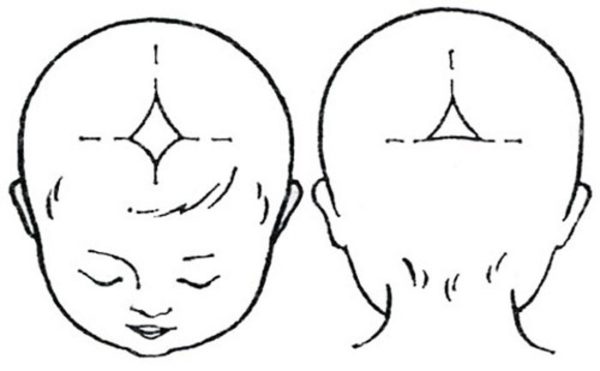
Where is
Determining where the fontanelle is located in a newborn baby is quite simple.
A large diamond-shaped fontanel measuring 2 by 2 centimeters is located right in the middle of the crown, or, as they usually say, on the top of the head.
A small fontanel is located on the back of the head. Its size is approximately half a centimeter.
When it overgrows
The large fontanel is overgrown by about the age of one year, sometimes there are slight deviations from this parameter until about one and a half years. But if the child meets age standards in other respects, then there is no reason to worry.
The small fontanel in children born at term is already closed. However, it happens that it was discovered after childbirth. Then its closure should be expected in two to three months.
The speed and time of closure of the fontanelles mainly depends on how much calcium the baby’s body is provided with. If there were no deviations in the mother’s diet and the optimal regimen for taking multivitamins was followed, then the overgrowth of the fontanelles usually occurs normally.
Developmental disorders
Knowing the timing when the fontanelle closes up, as well as its size, you can see any deviations, avoid and prevent the development of many dangerous diseases in newborns. Among them are several:
- Rickets . This disease is almost the most common cause of late closure of the fontanel. As a rule, this occurs in premature babies who are rarely exposed to the sun and have a lack of calcium and vitamin D. Read the article about rickets >>>;
- Hypothyroidism. A decrease in the amount of thyroid hormones may also cause a slowdown in the process of overgrowth of the fontanel;
- Down syndrome. Too large a fontanelle indicates the presence of this disease along with other characteristic signs;
- Overgrowth of the fontanel ahead of schedule may indicate excess calcium , and also indicate diseases such as craniostenosis, microcephaly;
- A depressed fontanelle is also a serious symptom. This phenomenon indicates acute dehydration of the body.
(picture clickable)
What does a too large fontanel or a slow (late closure) fontanel mean?
A careful examination of the child by specialists, a detailed description by the parents of the baby’s condition will be the key to early detection of abnormalities and will contribute to the correct prescription of preventive treatment.
Reasons for early closure of the fontanel
The fontanel is too small or the fontanel closes too quickly
Protruding fontanel?
Most often, a protruding fontanel is observed against the background of diseases that are accompanied by increased intracranial pressure: meningitis, encephalitis, tumors, intracranial bleeding, increased intracranial pressure for another reason.
If a bulging fontanelle is combined with one or more of the following symptoms, you should call a doctor as soon as possible:
- High fever;
- The bulging fontanel occurred after a head injury or a fall of the child;
- Vomit;
- Drowsiness or excessive irritability of the child;
- Strabismus;
- Convulsions or epileptic seizures;
- Loss of consciousness;
- Protrusion of the fontanel for a long time without other symptoms.
Sunken fontanelle?
Most often, retraction of the fontanel is observed due to dehydration of the child against the background of fever, diarrhea, and repeated vomiting. If you notice a sunken fontanel, make sure your child drinks plenty of fluids and contact a doctor to treat the illness that is causing the dehydration.
Fear of damage
Many people are very afraid of somehow damaging the fontanel. Remember! - this is practically impossible. Despite the apparent softness of the fontanel, it is very durable, and it cannot be damaged by ordinary manipulations (washing, bathing, combing, etc.).
Young parents often do not know why a thick film is needed on certain areas of the newborn’s head. The greatest fear is associated with the possibility of brain damage through the fontanel.
Find out the opinion of pediatricians about the age at which the fontanel should close, listen to the recommendations, and put aside fears and prejudices. Nature is wise: even such an incomprehensible formation on the head of a newborn brings benefits to the baby.
- general information
- Why do you need a fontanel?
- Norm and pathology
- When it overgrows
- Premature ossification
- It doesn’t take long for the “window” to close up
- Bulging and retraction
- Insufficient tension (sinks)
- Excessive tension (bulges)
general information
At the junction of the three bones of the skull after birth, a small “window” covered with dense connective tissue is felt. Many mothers are afraid that if accidentally pressed, the film will tear and the brain will be damaged.
The fears are unfounded: the surface of the fontanel is elastic, durable, and with careful touching, combing, kissing, and during bathing, it is impossible to damage the connective tissue. Gradually, the dense membrane ossifies, starting from the edges, the “window” decreases and becomes overgrown.
How many fontanelles do newborns have? Many parents do not suspect that there are six fontanelles on the baby’s head:
- two are clearly visible - front and rear;
- the “window” at the back closes soon after birth or no later than the first month of life;
- The fontanelle takes the longest to feel in the upper part of the head: at the junction of the frontal bone and the two parietal bones.
How to rinse a child's nose at home? We have the answer!
Read about the causes and treatment of green snot in children at this address.
Why do you need a fontanel?
Useful education performs several important functions:
- the role of a shock absorber for the skull during childbirth. When moving along the birth canal, the tiny head experiences strong pressure and becomes deformed (stretched). In the absence of soft but elastic connective tissue between the bones of the skull, the head would simply be damaged. Thanks to the elastic “windows”, the head again takes the correct shape, there are no signs of compression;
- role of thermostat in early life. A high temperature in a baby has a negative effect on the brain membrane, swelling and convulsions appear. The brain tissue is cooled through the connecting membrane;
- The “window” allows the brain to take on the desired size. In the first year of life, growth occurs rapidly; with a constant volume of the cranium, problems would arise with placing a larger volume of the brain inside a durable skull. The elastic membrane ensures a soft, smooth growth process.
Note! Elastic connective tissue softens the effects of falls. The baby will make a lot of “flights” while he masters motor skills. A durable, elastic membrane absorbs shock and prevents strong bone vibrations.
Norm and pathology
Most parents are interested in what the correct size of the “window” located on the crown of the head should be. Pediatricians consider the acceptable norm for a fontanelle in newborns to be 0.6–3.6 cm, sometimes a little more, but this is an exception to the rule.
At first, the “window” both decreases and increases slightly. This is not dangerous, the reason is simple: the fontanel becomes a little larger to allow normal growth of the brain.
When it overgrows
There is no exact time period for ossification of the entire surface of the connecting membrane. Nature has decreed wisely: don’t worry in vain if you regularly visit your pediatrician. The doctor monitors the condition of the large “window”; if there are deviations, a specialist will definitely tell you that the baby needs to be checked by a neurologist.
When does the fontanelle close in a newborn? The approximate period for the fontanel to harden and finally heal is 6 months to a year and a half. Sometimes the process ends a little earlier or, on the contrary, is delayed.
When to sound the alarm? Contact your pediatrician in the following cases:
- the “window” closes too quickly; by a month or two the connective tissue has almost completely ossified;
- the baby is almost two years old, and on the crown of the head a piece of dense membrane is still visible instead of bone;
- along the edges of the fontanel, the tissue does not stiffen, but on the contrary, becomes softer. If the edges are pliable, this fact often indicates the development of rickets.
Premature ossification
Causes:
- excess calcium and phosphorus during pregnancy. How to prevent side effects? While expecting your baby, follow the daily requirement of minerals and do not exceed the duration of the course of vitamin therapy prescribed by the gynecologist. The second recommendation is that if the connective membrane is rapidly ossifying, limit the amount of calcium;
- increased intracranial pressure. Sometimes the cranial cartilaginous sutures heal too quickly. The cause of the problem is hypertensive-hydrocephalic syndrome, meningitis. Be sure to contact a neurologist. The doctor will suggest ways to solve the problem of high intracranial pressure.
It doesn’t take long for the “window” to close up
Causes:
- hydrocephalus syndrome;
- developing rickets;
- lack of vitamin D;
- lack of calcium, phosphorus;
- violation of metabolic processes;
- lack of sunlight.
Important! Never make a diagnosis yourself. It is harmful to panic and attribute non-existent illnesses to the baby. Contact your pediatrician and neurologist. Doctors will talk with the parents, find out the daily routine, the duration of walks, and the diet of the nursing mother. If the child is artificial, it is important to know what infant formula the baby receives. Only after analyzing various factors will the doctor make a diagnosis, prescribe medications, and therapeutic measures.
Bulging and retraction
In the absence of pathologies, a slight swelling of the connective tissue follows the contours of the skull. It is easy to feel the pulse in the midline.
If the baby tenses up and cries for a long time, the bulge increases slightly and the pulsation is felt more strongly. This is a physiological phenomenon, everything is fine.
There are two types of connecting membrane voltage. Bulging or retraction of tissue requires mandatory monitoring. Show your baby to the pediatrician, visit a pediatric neurologist.
Insufficient tension (sinks)
Excessive softness of the edge, significant retraction often occurs with severe diarrhea, vomiting, and dehydration. It is necessary to call an ambulance and urgent measures to restore the water-salt balance.
Before the medical team arrives, be sure to give the baby a drink: every 5–10 minutes, give the child a teaspoon of boiled water.
Find out effective methods for treating caries of primary teeth in young children.
Medicines for inhalation with a nebulizer for a runny nose are described on this page.
Read here about how to teach your child to hold his head up on his own.
Excessive tension (bulges)
Connective tissue tension, weak arterial pulsation or its absence at rest indicate a possible increase in intracranial pressure. Another problem is added - the volume of cerebrospinal fluid increases in the spaces of the brain. If hypertensive-hydrocephalic syndrome is suspected, consultation with a neurologist is required.
Causes of the problem:
- difficult pregnancy and childbirth;
- excess vitamin D;
- inflammation of the meninges (meningitis).
Helpful Tips:
- when pronounced symptoms appear, pay more attention to the baby, think about what could cause the negative symptoms;
- if you develop vomiting, excessive drowsiness/excitability, unreasonable crying, refusal of milk in combination with suspicious phenomena on the surface of the fontanel, immediately call an ambulance;
- dehydration and very high intracranial pressure threaten dangerous complications for a small organism. Delay and attempts at self-medication are dangerous to the health and life of the little man.
Only a doctor will correctly assess the condition of the baby’s fontanel. Parents should carefully monitor the surface of the connecting membrane and control the density with light, soft touches. In case of deviations from the norm, severe bulging or retraction of the “window” on the baby’s head, consult a doctor in a timely manner.
More useful information about fontanel in newborns in the following video:
When it's dangerous
All of the above reasons are quite serious and have their own serious consequences, as a result of which the general condition of the child can be disrupted and the pace of his development can slow down. If the size of a large fontanelle in a newborn exceeds the permissible 3.5 cm, parents and the attending physician need to carefully monitor its condition and take daily measurements, including head circumference.

A prognostically bad sign is the constant bulging of a previously enlarged crown. This symptom most often indicates intracranial hypertension, which requires mandatory consultation with a neurologist and adequate therapy. In some cases, short-term bulging of the fontanelle is possible due to loud crying or screaming of the child, which does not require any diagnosis.
Quite often, with a long course of intestinal infection in an infant, one can detect retraction of the anterior fontanel. The appearance of such a sign indicates dehydration, which should be immediately relieved by administering fluids by mouth and infusion.
Rules for caring for a large fontanel
The large size of the crown requires proper care. Parents are advised to avoid putting excessive pressure on this area and try to move the child into different positions so that he is not in one position for a long time.
The possibility of injury, abrasions or scratches should be excluded. If peeling and yellow crusts appear in the fontanel area, they must be carefully combed out and the skin treated with baby moisturizing lotion. It is also advisable to avoid prolonged exposure to direct sunlight.
Thus, the fontanelles in a baby perform a number of necessary functions, and any pathological change in their size may indicate the presence of a disease. Therefore, both the mother and the pediatrician must monitor them until closure.
Properties of fontanel
The baby’s fontanel performs several important properties:
- Since not all parts of the skull bones ossify, they compress when the baby is born, and this makes it easier for him to pass through the birth canal. In this process, injuries are minimal for both the baby and his mother.
- The fontanel is important not only for easy childbirth, it is also necessary after the baby is born. Since the bones of his skull remain relatively mobile, and the growth and development of the brain occurs at a very rapid pace, the bone plates can adapt to the increase in its volume over the course of several more months.
- An important function of the fontanelles is cooling the brain, since the thermoregulation system in newborns is not yet sufficiently imperfect. Therefore, heat exchange occurs due to the movable spaces between the bones of the skull.
- If the baby accidentally falls, these same formations play the role of shock absorbers and reduce the likelihood of getting a concussion.