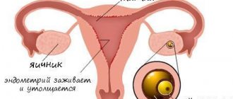
An embryo develops in the uterus in early pregnancy. It is protected by a thin film of the fertilized egg - the chorion, which during normal development is transformed into the placenta.
If during this period there is a failure in the development of the embryo, the embryonic egg is separated from its chorion film. The detachment site fills with blood. This pathology is called retrochorial hematoma during pregnancy. It can cause fetal development to stop and be lost.
What is retrochorial hematoma
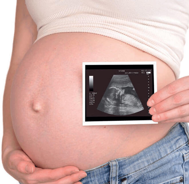
Ultrasound can detect hematoma during pregnancy. In the photo taken by an ultrasound diagnostic device, the characteristic darkening, as well as the deformation of the fertilized egg, is clearly visible.
A retrochorial hematoma develops during a singleton pregnancy at 5-7 weeks and in cases where a woman is pregnant with twins. Pathology can be suspected by the appearance of nagging pain in the lower abdomen, brownish or scarlet discharge. If a woman discovers these symptoms, she should immediately consult a doctor.
In addition to retrochorionic pathology, there are:
- subchorionic - it poses a serious threat to the fetus, surrounding the placenta;
- retroamniotic - timely detected formation, subject to adequate therapy, does not pose a threat to the developing baby;
- retroplacental - it is formed after the formation of the placenta, after 16 weeks of pregnancy;
- subamniotic - its presence is not dangerous for the baby, but requires regular monitoring;
- intrauterine - the presence of such a hematoma carries a serious risk to the fetus and requires urgent treatment.
Types of hematoma in the uterus during pregnancy
There are two types of hematoma. These are retrochorial and retroplacental.
Retrochorial hematoma
Retrochorial hematoma is the result of detachment of the ovum. In this case, there is a violation of the integrity of the small vascular network. The entire area that has been rejected is filled with blood masses. Pathology can be detected before mid-pregnancy.
This deviation is typical for ladies who:
- carry a child at a late age, after the 35-year barrier;
- have a history of thrombophilia;
- suffer from high blood pressure;
- got hit in the stomach;
- walking with multiple pregnancies;
- have a large amount of amniotic fluid.
Signs of retrochorial hematomas:
- pain in the abdominal area;
- slight brown stain on linen.
If the deviation is diagnosed in a timely manner, it will not have a negative impact on the course of pregnancy.
Retroplacental hematoma
This species is also detected in the second half of pregnancy, when the placenta is already fully formed. Here the cause of the pathology will usually be other diseases.
Danger for the expectant mother and baby
If a dangerous condition is detected on time, the woman has a great chance of recovering and saving the baby. With the normal further course of pregnancy and adequate therapy, the blood clot that has collected behind the membrane resolves within a month and is not detected on a subsequent ultrasound. This applies to pathologies whose blood clot volume does not exceed twenty milliliters.
The greatest danger comes from retroamniotic hematoma, or retroamniotic hematoma, as it is called. This is hemorrhage between the amniotic sac and the wall of the uterus. It is dangerous because it is accompanied by bleeding and threatens the mother with blood loss.
Any unpleasant sensations in the lower abdomen or spotting during pregnancy require attention from the woman and specialists. They may be a sign of retrothecal hematoma. To diagnose the condition, a blood test is ordered and one or more ultrasound examinations are performed.
If the hemorrhage occupies about half the surface of the separation of the uterine wall and the chorion, and the volume of blood accumulated in it exceeds twenty milliliters, there is a high probability of a frozen pregnancy.
Even if the disease was stopped at an early stage and the blood clot has resolved, the risk to the fetus still remains. The subsequent formation of placental insufficiency, its premature “aging”, a reduction in the amount of nutrients supplied to the baby, the development of hypoxia, as well as the birth of a low-weight baby are possible.
Danger of hematoma during pregnancy
If there is a detachment of the fertilized egg, then there is a risk:
- the occurrence of bleeding;
- inhibition in embryo development;
- miscarriage or premature birth.
In order for everything to continue to proceed normally, treatment should be started at the initial stage.
Does it always appear with discharge?
Hematomas on the uterus may not manifest themselves as any bruising. Bleeding can only be detected by instrumental examination - ultrasound.
What type of hematoma is noted without discharge?
A retroplacental hematoma may not be noticeable by discharge. In this case, the woman feels only pain in the abdominal area.
Pain symptoms
Pain in women who have been diagnosed with a hematoma on the uterus is usually present in the abdominal area, from below.
There is often discomfort in the lumbar region.
The pain is nagging and may throb. They intensify during physical activity, after physical labor.
Why does the disease occur?
Causes of retrograde hematoma:
- a change in hormonal levels in a woman’s body, provoked by bearing a baby;
- infectious diseases of the genitourinary system;
- severe toxicosis of pregnant women;
- blood pressure surges, hypertension;
- severe nervous stress;
- disruption of metabolic processes in the body;
- the presence of pathologies in the uterine cavity;
- addiction to nicotine and alcohol;
- increased physical activity;
- the presence of a benign or malignant tumor in the expectant mother;
- autoimmune diseases;
- blood clotting disorder;
- overweight.
Features by trimester
Placental abruption during pregnancy can occur in any of the three trimesters; each of these periods has its own clinical characteristics, as well as the degree of danger for the mother and her unborn child.
- First trimester. Placental abruption in the early stages of pregnancy in the first three months (weeks 1-12) occurs frequently, but it can be prevented with timely diagnosis and proper treatment. The degree of complexity of the situation helps to clarify the ultrasound, which fully reveals the picture of the processes occurring in the woman’s body. If the situation can be stabilized, the pregnancy continues and its further development proceeds normally.
- The second trimester (up to 27 weeks) is characterized by dizziness, severe weakness, high tone of the uterus - it becomes very tense and painful. If placental abruption occurs in the second trimester, the baby stops receiving the required amount of oxygen, which usually comes in the bloodstream from the mother’s body (at the same time, the pregnant woman feels violent movements, much more intense than usual). This condition can be accompanied by bleeding of various types and intensity. Increasing hypoxia can only be stopped by an urgent caesarean section.
- Third trimester. The most common occurrence of detachment is in the later stages - in the third trimester. During the period from 28 to 36 weeks, the symptoms of the pathology look classic - pain, bleeding, signs of fetal hypoxia. A feature of the period is the most dangerous premature placental abruption, which poses a threat not only to the baby, but also to the mother. The uterus becomes so oversaturated with blood that it loses the ability to contract. In this case, heavy blood loss cannot be stopped; to save the woman, the organ is removed. If such events occur in the last month of pregnancy, tragedy can be avoided if childbirth is carried out in a timely manner for medical reasons. However, if placental abruption has occurred only partially and there is no bleeding, pregnancy continues until the physiological time of labor.
How to identify a hematoma - symptoms of the disease
Symptoms of the disease depend on the severity of the disease, its nature and stage. If the pathological changes are mild, the expectant mother may not be aware of their presence. They are detected only with the help of ultrasound diagnostics. Bloody discharge does not appear because blood cells are retained by the chorionic villi. This pathological condition passes without threatening either the pregnant woman or her baby.
The average degree of the disease is manifested by nagging pain in the lower abdomen and lower back, as well as brown discharge. If the color of the discharge is brown, the woman’s condition does not worsen, and the discharge does not change its color to scarlet, specialists are in no hurry to resort to treatment. This condition indicates that the retrochorial formation is in the stage of resorption, that is, it comes out on its own.
The severe form of retrocapsular hematoma is characterized by nagging pain in the lower abdomen with cramping attacks. The condition is accompanied by a decrease in blood pressure, bleeding begins. Loss of consciousness is possible. Emergency medical care and immediate hospitalization are required.
Hematoma of the fertilized egg causes
Pregnancy is a wonderful time in the life of every woman. The birth of a new life, bright emotions and feelings overwhelm the expectant mother. Unfortunately, sometimes this unforgettable time is overshadowed by health problems. One of the dangers may be a hematoma in the uterus. After all, the uterus experiences enormous stress during pregnancy. Uterine hematoma (from the Greek “heme” - “blood”) is a space filled with blood from damaged blood vessels. There are several types of hematomas.
Retrochorial hematoma during pregnancy. What it is?
This is an accumulation of blood between the wall of the uterus and the chorion.
The chorion is the outer villous membrane that surrounds the embryo in the uterus. Its main functions are protection against infections and supplying oxygen to the embryo. The chorion is covered with many villi. In the future, the placenta will form from it. Sometimes it is even called the “early placenta,” which indicates its importance for the normal development of the embryo. A retrochorial hematoma during pregnancy indicates that the chorion began to exfoliate, a space appeared between it and the wall of the uterus, and it filled with blood. This diagnosis can only be made before the 16th week of pregnancy.
Due to the fact that the hematoma is located between the membrane of the fetal egg and the uterus, retrochorial hematoma during pregnancy is also called transthecal or retrothecal.
Of course, such detachment should not normally occur. The consequences can be very sad - the hematoma puts pressure on the amniotic sac, interferes with normal blood supply, contributes to the development of nutritional deficiencies and can lead to miscarriage. Even if a miscarriage does not occur, the consequences of a hematoma can affect the development of the unborn child's brain. Indeed, with this pathology, oxygen starvation of the embryo often occurs. And as you know, it primarily affects brain cells. Therefore, you should not neglect the doctor’s prescriptions, even if a woman has no discharge or pain with a retrochorial hematoma.
The causes of retrochorial hematoma have not been fully established. But the likelihood of its occurrence is increased if there are:
1. malformations or insufficient development of the uterus and other genital organs;
2.hormonal imbalances;
3. infectious or autoimmune diseases;
4.excessive physical activity;
5.abdominal injuries or bruises;
6. any adverse environmental influences - noise, vibration, environmental unfavorable factors;
7. disorders in the blood coagulation system;
8.psychological tension, stress;
9.anomalies in the development of the fertilized egg or embryo;
10.bad habits of a pregnant woman
11.individual characteristics of the body (including hereditary predisposition).
Unfortunately, it is often difficult to determine the true cause of the anomaly. Therefore, a pregnant woman should be attentive to her health, avoid overwork, heavy physical work and negative emotions.
Most often, retrochorial hematoma is discovered by chance, during a routine ultrasound at 12 weeks of pregnancy, since there are no external manifestations. The woman is not bothered by anything; there is no unusual vaginal discharge. Less commonly, a pregnant woman complains of nagging pain in the lower abdomen or lower back, and is bothered by weakness and spotting. Often retrochorial hematoma occurs at 5–7 weeks of pregnancy.
If the pathology is severe, the woman is bothered by brownish discharge mixed with scarlet blood, nagging, cramping pain in the lower abdomen. Due to blood loss, blood pressure may drop, chills, weakness, and dizziness may appear. In this case, you must urgently seek qualified medical help. But such cases, fortunately, are quite rare in modern medicine.
Of course, upon hearing about the hematoma, the expectant mother begins to worry, because this may indicate a threat of termination of pregnancy. However, it is best to try to pull yourself together, as mom’s nervousness will only worsen the situation. It should be remembered that most often this pathology can be successfully treated. The sooner treatment is started, the greater the likelihood that the pregnancy will continue to develop successfully. Much depends on the size of the hematoma. Of course, the smaller it is, the better. Hematomas that do not exceed 40% of the size of the gestational sac are best treated. In any case, the main condition for the resorption of a hematoma is rest, and ideally, bed rest. In addition, the doctor will conduct an examination of the pregnant woman and a medical examination in order to find out the possible cause of the pathology and begin its drug treatment. If the doctor advises you to continue it in a hospital setting, you should not refuse.
It should be noted that if a pregnant woman was diagnosed with a retrochorial hematoma, and after some time she began to have brownish discharge, then, most likely, the hematoma has resolved, and this is the “old” blood with which it was filled. It is not precisely established how long it will take for the hematoma to “come out.” Presumably - about 2-5 weeks. But scarlet vaginal discharge is an alarming symptom. Most likely, the hematoma continues to grow - and there is a threat of spontaneous termination of pregnancy, that is, miscarriage. It is best to go to your doctor, who will conduct an examination and make an accurate diagnosis.
If an ultrasound was performed and the doctor wrote in the conclusion that the retrochorial hematoma is at the stage of organization - this means that you can exhale - the threat is over! Now all that remains is to be patient and wait for it to either resolve or come out with vaginal discharge.
Another type of diagnosis that a pregnant woman may hear about is retroamniotic hematoma. What is retroamniotic hematoma during pregnancy? It is a hemorrhage between the wall of the uterus and the amnion. Amnion - amniotic sac, or popularly known as “shirt”, is a kind of sac that contains the fetus and amniotic fluid. The amniotic membrane is very thin, but elastic. Its main function is to protect the child and establish a relationship with the mother’s body.
The causes of retroamniotic hematoma are the same as retrochorial hematomas. However, retroamniotic is less dangerous for the fetus, since it is protected by the amniotic sac. To a greater extent, this hematoma is dangerous for the mother, as it is accompanied by bleeding. The most important thing is to detect it in time and begin treatment. Every tenth pregnant woman experiences a hematoma during pregnancy, and only 2-3% have an unfavorable outcome and end in miscarriage. In other cases, pregnancy proceeds normally. There are even cases when the doctor learns about the existence of a hematoma after childbirth, seeing its traces on the placenta. That is, a hematoma does not always pose a danger to the child.
The attending physician, based on knowledge of the nature of the hematoma and its size, can recommend delivery by cesarean section - this eliminates hypoxia, that is, oxygen starvation of the child during childbirth.
The main treatment for various types of hematomas during pregnancy is to prevent its growth, which means it is necessary to exclude harmful external effects on the pregnant woman’s body. The expectant mother should take care of her health - give up bad habits: do not drink alcohol even in small doses, avoid smoking and taking medications. Of course, there are cases, especially in the early stages of pregnancy, when a woman does not suspect that she is pregnant and continues to lead her previous lifestyle. However, having learned about the birth of a new life, it is necessary to urgently consult a doctor and inform about the medications you are taking. Doctors also advise pregnant women to take greater care of their health from colds and infections - if possible, avoid drafts and crowds of people. However, it also happens that a woman was a “model” pregnant woman, followed all the rules, but developed a hematoma. In this case, all that remains is to be patient and tune in to a positive outcome.
Tumor treatment
Treatment of retrotracheal hematoma is carried out under the strict supervision of an obstetrician-gynecologist. His tactics come down to two directions: stopping bleeding, blocking tumor growth and its gradual resorption. At all stages of treatment of the disease, the use of magnesium drugs, antispasmodics (Nosh-Pa), which relieve uterine tone, as well as hemostatic agents, if necessary, is indicated (Ascorutin, Dicynon).
Often, the doctor prescribes mild sedatives (tincture of valerian) to stabilize the emotional state of the expectant mother. Patients are prescribed medications that improve uteroplacental permeability, as well as some homeopathic remedies. The form and dosage of all drugs is determined by the doctor depending on the severity of the disease.

Mild forms of the tumor can be treated at home, but under the supervision of a doctor, without missing scheduled specialist appointments and mandatory tests. Inpatient treatment is preferable, since at home it is more difficult for a woman to comply with all instructions, especially adherence to bed rest.
The expectant mother must do a blood test throughout the entire period of bearing the baby to monitor fibrinogen levels. The severe course of the disease requires mandatory hospitalization.
During treatment, a woman must remain in bed, avoid physical activity, stress, and adhere to a healthy lifestyle. Working women are entitled to sick leave during treatment. The duration of therapy is determined by the severity of the condition. On average it ranges from one to three weeks.
Will a caesarean section be required?
The indication for delivery by cesarean section is a retroplacental formation that occurs in the later stages. If the diagnostic results indicate that the fetus is suffering from its presence, the operation is scheduled a little earlier than the expected date of natural birth.
How does a hematoma heal?
The fact that the hemorrhage is resolving is indicated by spotting or moderate brown discharge from the vagina. How long it takes for a retrochorial formation to emerge depends on the timing of its appearance and the size of the blood clot. On average, discharge lasts from 15 to 35 days. It is important to make sure that the appearance of discharge does not mean a missed pregnancy. The gynecologist who monitors the pregnancy and an ultrasound diagnostic specialist will tell you exactly how long the tumor will resolve.
Can a hematoma not come out?
The structure of the female pelvic organs in some cases does not allow the tumor to come out in the form of brown discharge. This is impossible when it is located high at the fundus of the uterus. In such cases, it slowly resolves without the threat of termination of pregnancy. You can read about this on pregnancy forums and read numerous reviews from those who had such a hematoma.
Consequences of the disease
From the moment a blood clot appears until delivery, the woman is under close medical supervision. If she strictly follows all the recommendations, the prognosis is positive. In most cases, with adequate and timely treatment, the blood clot comes out on its own or resolves without harming the growing baby.
Without timely medical assistance, a retrothecal hematoma during pregnancy leads to:
- intrauterine fetal death;
- uterine bleeding;
- miscarriage;
- abnormalities in child development;
- placental abruption at 8-9 months.
If no measures are taken, the bleeding will not stop, which means the tumor will increase in size, threatening the safety and normal course of the gestation period.
Urgent dangerous conditions
The most dangerous situation and requiring immediate hospitalization is considered to be a situation in which a hematoma on the uterus formed in the first trimester of pregnancy and has already reached a large size (a quarter of the size of the entire organ).
In the event of such a deviation from the norm, a woman may have a miscarriage at any time. Here any doctor will say that in this state there is intrauterine growth retardation of the fetus. Hypoxia is noted, and increased uterine tone is present.
In an inpatient setting, the woman will be prescribed effective treatment to normalize blood flow. Your health will improve.
Intelligent treatment can prevent miscarriage even at 6-8 weeks, which is why it is important to go to the hospital in the early stages and do everything necessary, as the doctor says.
Uterine hematomas are retrochorial and retroplacental. They have a common symptom in the form of nagging pain in the lower abdomen. Any doctor who is competent in the field of gynecology and conducts ultrasound diagnostics of pregnant women can diagnose the disease. Bruises will be clearly visible on the screen, indicating detachment of the fertilized egg. It is important to start treatment immediately. So that the pathology does not increase in size and does not affect the development of the fetus and the course of pregnancy.
How to prevent hematoma - preventive measures
The appearance of this unpleasant complication can be avoided if the expectant mother leads a healthy lifestyle and is attentive to her new condition.
Main preventive measures:
- Rejection of bad habits. If a woman smoked before conception, then during the period of bearing the baby and feeding him, you should forget about smoking.
- Timely treatment of infectious viral diseases. Gestation is not the time to experiment with your health.
- Maximum compliance with the doctor’s instructions if there is a risk of developing retrochorial pathology. In this case, you need to stay in bed and avoid physical and emotional stress. It is recommended to lie with a bolster or pillow under the pelvic area to drain the blood.
- Avoid bruises, injuries and falls.
- Do not lift or carry heavy objects.
- Monitor your diet. It is necessary to consume foods rich in vitamins that stabilize the functioning of the gastrointestinal tract and cardiovascular system. It is advisable to exclude foods that cause constipation and gas formation in the intestines.
- Do not delay registration. The earlier the pathology is identified, the easier it will be to overcome it without consequences.
Watch an informative video on the topic of miscarriage:
Determination of hematoma in the uterus
Uterine hematoma can be detected using ultrasound. On the screen, the specialist will see all the deformations present in the fertilized egg, the thickness of the uterine walls and all associated complications.
To clarify and conduct a detailed study of the condition of the organ, the woman is referred to:
- general blood and urine tests;
- coagulogram;
- dopplerometry;
- hormonal diagnostics.
Urine and blood tests
General tests will help you see if there is an infection or inflammatory process in the body. Such laboratory tests are advisable and mandatory throughout gestation, every 2-3 weeks.
What does it look like on an ultrasound?
Below we present to your attention an ultrasound scan showing a hematoma on the uterus.
Coagulogram
A coagulogram means a blood test that determines its coagulability. Diseases of the gastrointestinal tract, liver, and the very “interesting” position in which the woman finds herself can change the indicators for the worse.
Smear and detection of infections
To rule out infectious diseases on the genitals, the gynecologist will analyze the discharge. It is taken from the vaginal area - a smear. Results can be obtained after 1-3 days, depending on the clinic.
Doppler
This diagnostic procedure is carried out using a special device. The ultrasound wave will display an image on the screen that will indicate the presence of blood flow in a particular place. The specialist records the speed of blood flow, the diameter of the vessels and determines the pressure in the vascular lumens. In addition to the condition of the placenta, using Doppler, you can find out what condition the heart and blood vessels of the fetus are in, and whether there are any abnormalities. You can determine hypoxia, intrauterine infection, and whether there are umbilical cord loops near the baby’s neck.
Hormonal diagnostics
They may also be asked to donate blood for hormones. Venous blood is taken. If there is a deficiency or excess of any hormones, then this may be the cause of the hematoma.