How is pregnancy calculated?
Doctors use two methods for calculating gestational age: obstetric and embryonic. The first option is most often used. The first day of pregnancy is the beginning of menstruation.
Normal intrauterine development of the fetus lasts about 40 weeks. In this case, an error of 14 days or less is allowed. If the birth of the baby occurred much earlier, then we can talk about premature birth. The onset of contractions and opening of the cervical canal in the first half of pregnancy leads to miscarriage. The period of development of a child in the womb can be determined using manual examination or ultrasound diagnostics. Moreover, the second method is more reliable and reliable.
First week of obstetric period
The concept of the first obstetric week is very conditional. The calculation is carried out from the first day of the missed period, because this date is most often remembered by patients.
However, the exact day of conception is quite difficult to determine. Even if this is an established married couple who clearly remembers the date when they had unprotected intercourse, then conception itself may not have occurred on that day. Fertilization of the egg can occur on the third day after sexual intercourse.
Obstetric period is calculated from the last menstrual cycle. The embryonic period is the fertilization of the egg itself. Moreover, the first period is longer than the second by about two or three weeks. This is explained by the fact that a woman takes 14-18 days from the first days of her period to ovulation (at this time she becomes pregnant). It turns out that the first week of pregnancy according to the obstetric period is not such.
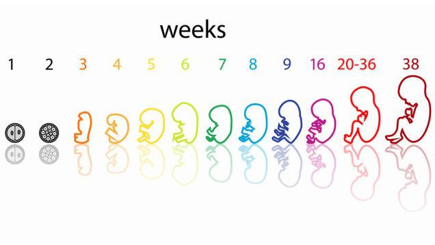
How does a child develop during the weeks of pregnancy?
During the period from the first to the forty-second week, a colossal transformation of the fetus occurs. From a few cells that are not visible to the normal eye, the embryo develops into a real little person, capable of life outside the mother's body. Doctors closely monitor the growth of the baby in the woman’s belly. For this purpose, regular inspections, measurements and weighings are carried out. Based on this, doctors make a conclusion about whether the fetus is developing correctly.
The development of the baby can be tracked by week of pregnancy using ultrasound. In this case, doctors have certain standards with which the data obtained are compared. Based on such a study, we can conclude whether there is a delay in fetal development or whether the pregnancy is proceeding normally. Let's take a closer look at how the baby grows and changes with each subsequent week.
Pregnancy calendar by week
The pregnancy calendar by week is very convenient. It will facilitate medical supervision of the expectant mother and her baby, and will also give answers to the most important questions that will certainly arise in front of you: when the belly begins to grow, why does one feel sick during pregnancy, how to recognize the first movement of the fetus.
Thanks to this calendar, you will not forget that the time has come to undergo screening, take tests, or go for a routine ultrasound. And as the due date approaches, such a weekly calendar will help you best prepare for the long-awaited event.
First month of pregnancy
All stages of fetal development are divided into trimesters. There are three of them in total. The first trimester is already in full swing when a woman receives a positive pregnancy test result. The countdown begins long before this.
The first month of pregnancy includes the first, second, third and fourth weeks. From days 1 to 5-7, a woman continues to menstruate. However, during this period there is already an active production of hormones that affect the ovaries and provoke the growth of follicles. From days 8 to 14, the dominant phyllicle is released and the endometrium grows for embryo implantation. The third week is characterized by the release of the egg from the follicle and its fusion with the sperm. Conception has already taken place, but the woman is not yet aware of it. From the 22nd to the 28th day, the fertilized egg continues to actively divide and move through the fallopian tubes towards its goal. When a set of cells reaches the reproductive organ, secure attachment occurs. This is where the pregnancy will progress.
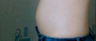
With the onset of a delay in menstruation, a woman may become suspicious of her new situation. However, to confirm her suspicions, she needs to conduct home testing or take a blood test.
Visiting the doctor at 4 weeks pregnant
There are several ways to confirm pregnancy. First, the pharmacy test will show two stripes. The second way is to wait for the expected date of the new menstruation and make sure that there are no periods, that is, a delay. The third way is to do an ultrasound examination at the end of the fourth obstetric week, in which you can see the embryo (small dot) on the surface of the endometrium of the uterus. A photo at the 4th week of pregnancy (at the end of the 4th week - the beginning of the 5th) will clearly indicate the fact of an “interesting situation”. The fourth method is to take a blood test for hCG.
You should go to a doctor for a consultation only if the fact of an “interesting situation” is confirmed by one of the indicated methods.
In the fourth obstetric week, it is still too early to register with a local obstetrician-gynecologist at the antenatal clinic at your place of residence. This can be done up to the twelfth week.
During your initial visit, the doctor will conduct an external examination, collect your data, recommend multivitamin complexes for pregnant women, give lifestyle advice and prescribe the necessary tests and examinations for the next visit.
First trimester
Every expectant mother should know everything about the development of the child during the weeks of pregnancy. The first trimester lasts from the first to the thirteenth week. During this period, the embryo from several cells will turn into a small resemblance to a person. How does the child develop during the weeks of pregnancy during this period?
Week 5
At this stage, the woman is most likely already aware of her new position. The future baby's vital parts are actively developing: the respiratory tract, pancreas and liver. All this is quite difficult to examine using an ultrasound machine, but a competent specialist and good equipment are capable of this.
In the yolk sac, important cells - germ cells - are formed. If you are expecting a girl, then these are the eggs. For boys, these will be sperm. The fertilized egg increases in size and by the end of the week occupies about a quarter of the reproductive organ. The future baby looks like a small disk and has a size of no more than two millimeters.
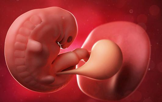
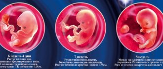
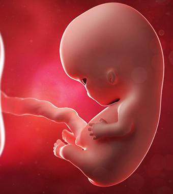
Week 6
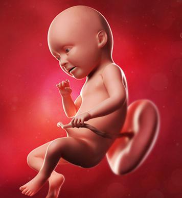
During this period, the child looks more like a fish: he has a small tail and gills. Don't worry - by the time you give birth, all this will disappear. The size of the baby from the sacrum to the head is about four millimeters. He floats freely in his bubble and has no restrictions. The child is connected to the mother with a thin tourniquet, which will later become the umbilical cord.
At week 6, a very important event occurs: the baby’s heart begins to beat. Outwardly, it looks more like a fast pulsation. However, with the help of a good ultrasound machine it is quite possible to count muscle contractions. Their average number reaches 160 beats per minute.
At this stage, the baby’s face is actively transforming. He already has side holes that will later become the ears, mouth and nose. In addition, the liver learns to secrete blood cells, and the rudiments of the brain monitor the heartbeat.
Week 7
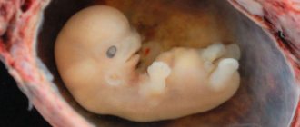
This week the baby is gaining a lot of growth. It doubles in size and is already 10-12 millimeters. It is worth recalling that this measurement is made from the head to the sacrum.
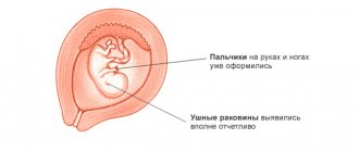
The baby's legs and arms take on a human shape. However, they are still more like flippers due to the presence of membranes between the toes. The intestines increase in size. The brain is divided into two parts - hemispheres. The heart of the unborn child protrudes strongly and already has several chambers. The growth and development of the fetus is actively occurring.
8 week
The growth of the embryo at this stage is from one and a half to two centimeters. The genitals are actively forming. During this period, they are the same in girls and boys, but soon the genital tubercle will turn into a penis or clitoris.
The baby's fingers increase in size and become human-like. The stomach passes into the peritoneum and acquires nerve endings there. It is these structures that will be needed in the future for normal digestion. At this stage, the heart is almost formed. It has a connection with large vessels and veins.
Week 9
At this time, the size of the baby is about three centimeters. You can already see during the ultrasound that the tail has disappeared. The weight of the embryo at this moment is about one gram (the weight of the child will actively increase over the weeks of pregnancy).
The heart muscle has split into four parts and is already functioning quite well. During this period, the rudiments of future teeth are laid. This is why the expectant mother may suffer from calcium deficiency. However, it should be used only if the doctor recommends it.
Week 10
During this period, the baby has a length of three to four centimeters. Its average weight is four grams. The period of formation of all important organs has come to an end.
The baby's arms and legs begin to move. They actively bend and unbend. All this can be seen during an ultrasound examination. The liver continues to actively secrete blood cells, the heart contracts, and the brain functions. The child’s body acquires a new covering – fluff. It will disappear shortly before birth. Marigolds and pads form on the fingers.
Week 11
The baby weighs approximately 7 grams. He is already making active movements, although the woman still does not feel it at all. The main parts of the body are strengthened, including the neck.
A special feature of this period is that the child may begin to hiccup. This is a consequence of the formation of the diaphragm. There are no leukocytes in the baby’s blood yet; this substance only contains red blood cells. The baby's legs and arms are becoming more and more proportional, but the upper limbs are still slightly longer.
Week 12
At this time, the length of the child from the buttocks to the head is about 6 centimeters. All previously formed organs continue to grow and develop. The child learns to perform new movements. He opens his mouth wide and tries to capture liquid. Eyes may close. The child's facial expressions are formed.
Week 13
At this time, the first trimester ends. Fetal development continues to progress. Weight is approximately 25-30 grams, and height from crown to buttocks is about 6-8 centimeters. An image appeared on the pads of my fingers. It is individual for each person.
If you are expecting a girl, her ovaries will develop this week. They are still very small, although they contain more than two million sex gametes. It is worth noting that by the time they are born there will only be a few hundred thousand left.
last weeks of pregnancy
Yes, I haven’t been here for a long time, although I have always loved writing. And then it turned out that I disappeared completely, although maybe my thoughts and experience would be useful to someone.
And the reason is this. When, at the third ultrasound, I found out that our baby was still not trying to turn over on her head, I, of course, was terribly upset, and I didn’t want to write about it at all. I kept waiting for it to turn over, sit down and write. In the meantime, I did all sorts of exercises that, according to doctors, contributed to the revolution, visualized, stood on my ears (in the literal practical sense). But the daughter, apparently, had her own plans for how she would come into this world.
In the last week before the New Year, my husband and I decided to ask the opinions of the doctors at the First Maternity Hospital on Vasilyevsky, where I planned to give birth. In the emergency room they quickly installed an ultrasound scanner and said that the baby was big and would not turn over on its own, and therefore you are welcome to have a cesarean section at 39 weeks. We began to look for specialists who would advise on the obstetric turn, but the holidays were already approaching, and until January 11 no one was ready to see us.
I cried a lot, stood, again, on my head, swam in the pool, hoped for a miracle and asked myself whether I was ready for a possible operation. On the one hand, what is there to prepare for: you come to the hospital, take tests, get anesthesia - or general anesthesia - and here you are, your baby. Minimal risk, no worries and dangers associated with breech birth, which, by the way, in the First Maternity Hospital, according to the head of medicine, they do not like to accept due to possible injuries.
But how can you give in to the operation and miss everything? This question was just driving me crazy. Planning a pregnancy, carrying a baby for 9 months, going to classes to prepare for childbirth with your husband, learning to breathe correctly... and all this in vain? In addition, I was confident that it was important for a child to be born on his own, well, the recovery after childbirth and the second scar on my stomach (the first remained from abdominal surgery 7 years ago) did not please me much.
Meanwhile, I was already 38 weeks pregnant. At the antenatal clinic, having learned that I wanted to give birth myself, they twirled their finger at my temple and wrote out a referral for hospitalization with an open date. I went to several appointments with an osteopath in the hope that maybe we could fix something that was bothering the baby. By the way, after these appointments I felt a lot better - before that I was so stiff that it was difficult to stand in an upright position. And she advised me to go to another maternity hospital, where there is a practice of delivering babies in a breech position.
There was hope again. On January 15, 10 days before giving birth, my husband and I went for an ultrasound to Maternity Hospital 17 in St. Petersburg, deciding that today we must make a decision, but only after a competent ultrasound. It so happened that we had an appointment with the head doctor, who did an ultrasound, examined everything and said that he saw no reason not to give birth herself. My pelvis is wide, the baby, as he said, is not huge, there are no entanglements, I have a positive attitude, so everything will work out. And an emergency caesarean section, if necessary, will always be done in time.
We left the office with a feeling of joy and enormous relief! It was decided to calm down, tune in for the best and calmly wait for day X.
Second trimester
During this period, the baby’s weight actively increases over the weeks of pregnancy. During an ultrasound examination, body length from the tailbone to the crown of the head is no longer measured. Experts often provide formulaic calculations and announce the approximate height of the baby. How does a baby grow in the second trimester? Let's look at the intrauterine development of the fetus week by week.
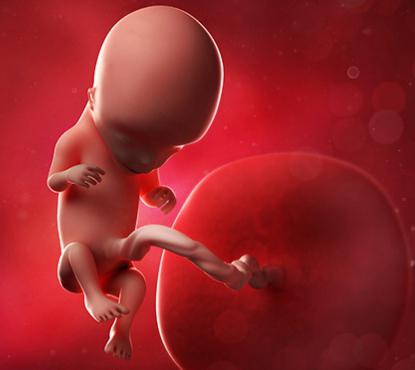
Week 14
The length of the baby is about 9 centimeters. The kidneys acquire a new function - urine production. The intestines become even longer and perform the first contractions.
The blood produces leukocytes - white blood cells that will protect the baby after birth. The process of formation of the vocal cords begins. You will hear the first sounds made by the baby immediately after its birth. Nerve endings are formed. The baby can already react to external stimuli and feel his mother’s touch on his stomach.
Week 15
The baby's height at this moment is from 9 to 10 centimeters. His eyes are still closed, but the baby can clearly distinguish light stimuli. The baby is actively moving, and the expectant mother at this stage can already feel light tremors.
The legs have grown and become longer than the upper limbs. The child is actively developing the respiratory system. It forcefully pushes amniotic fluid out of the lungs. Don't worry, this substance is absolutely sterile and can be renewed more than 10 times in 24 hours.
Week 16
The baby's weight at this moment is about 70-80 grams. The bladder is actively working, releasing fluid approximately once an hour. The intestines are gradually filled with contents that will be released in the first days after birth.
The stomach and gall bladder begin to function actively. The child already has his own blood type and Rh factor.
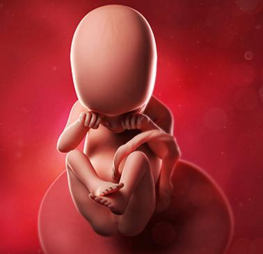
Week 17
The baby weighs approximately 100 grams. During this period, subcutaneous fatty tissue is formed. It will play an important role in metabolism.
The baby can control his movements. He brings his hands to his face and sucks his finger. The child closes his eyes with his palms when he doesn’t like something. Most of the processed substances are excreted through the placenta, despite the fact that the kidneys can already do their job quite well.
Week 18
The child weighs approximately 150 grams. Girls have already formed fallopian tubes and ovaries. These organs occupy the right place in the peritoneum. The boy's testicles have not yet descended into the scrotum. This can only happen after birth.
The child’s endocrine system begins to work actively. It is worth noting that if the mother has some problems, then the baby can help her body. However, such a replacement is fraught with disturbances in the child’s body. This is why it is so important for a mother to monitor her hormonal levels.
Week 19
The baby weighs approximately 200 grams. The first real hairs appear on his body, which are very different from the fluff. The brain is actively transforming and acquiring new functions. The baby can hear, taste the amniotic fluid, and acquires vision and touch.
Fatty lubricant forms on the baby's body. It protects the baby from bacteria and helps him easily pass through the birth canal.
Week 20
The weight of the baby is from 250 to 300 grams. Height is 23-27 centimeters. The child's heart becomes larger and beats faster. From this date, many doctors begin to measure contractions of this muscle using a special tube.
The excretory system is still actively working. There is already a certain amount of feces in the intestines - meconium. It is worth noting that it will only be released after childbirth. Preliminary bowel movement indicates oxygen starvation of the child and requires urgent medical attention.
Week 21
The weight of the baby is approximately 320-400 grams. From the body of the expectant mother through the placenta, it begins to receive immune cells. The vestibular apparatus actively develops and trains during movements.
Week 22
The height of the unborn baby is approximately 28 centimeters. The baby's weight is about 450 grams. Eyelashes begin to grow and eyebrow arches begin to form. The baby moves and pushes more and more.
Week 23
What characteristics does the fetus have during this period? 23 weeks of pregnancy is the period when the baby weighs approximately 500 grams. After overcoming this border, he has every chance of surviving premature birth. However, this will require certain equipment.
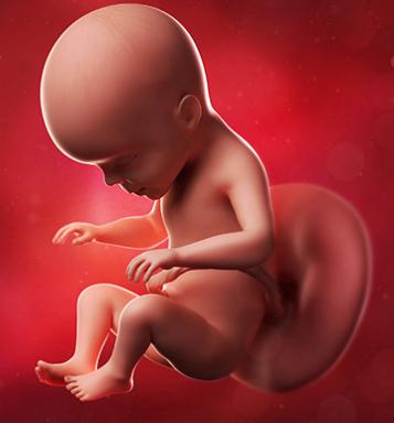
Week 24
During this period, the child’s weight has already exceeded half a kilogram and is approximately 600 grams. The baby's height is 28-32 centimeters. Most often the baby is in a state of sleep. At this time it is actively growing.
Week 25
The baby's weight is about 700 grams and his height is more than 30 centimeters. The bones and cartilage are strengthening every minute, but they are still very fragile and delicate.
Week 26
The baby's weight is approximately 750 grams, and its height exceeds 32 centimeters. During this period, the child's pituitary gland takes responsibility for the baby's growth. Previously, this directly depended on the mother’s body.
Third trimester
This period is characterized by strong growth of the baby. All organs and systems are already formed and function as they should. The norms of fetal development in the third trimester are as follows:
- week 27: 34 centimeters and 900 grams;
- week 28: 34.5 centimeters and 1050 grams;
- week 29: 36 centimeters and 1150 grams;
- week 30: 40 centimeters and 1300 grams;
- week 31: 41 centimeters and 1550 grams;
- week 32: 42 centimeters and 1700 grams;
- week 33: 43 centimeters and 1950 grams;
- week 34: 44 centimeters and 2200 grams;
- week 35: 45 centimeters and 2.5 kilograms;
- week 36: 46 centimeters and 2.6 kilograms;
- week 37: 49 centimeters and 3 kilograms;
- week 38: 49.5 centimeters and 3.03 kilograms;
- week 39: 50 centimeters and 3.2 kilograms;
- week 40: 53 centimeters and 3.5 kilograms.
Important dates in diagnosing a child’s condition
The 32nd week of pregnancy plays a big role. The baby’s weight and height allow us to draw a conclusion about what size the baby will reach by the time of birth. During this period, a final ultrasound examination is performed.
The timing of fetal development is also taken into account when ordering the first ultrasound. More often, doctors choose the period from 11 to 13 weeks. At this time, the presence of some congenital diseases and syndromes can be detected.
Although the 32nd week of pregnancy is very important, the baby’s weight also plays a big role at 20 weeks. During this period, doctors can detect whether there is intrauterine growth retardation.
Possible deviations
In some cases, during an ultrasound examination it turns out that the baby is very small or large. Often this is a consequence of an incorrectly set period, but it can also be a sign of certain diseases. Don't panic ahead of time. In this case, doctors prescribe correction with certain medications and recommend repeating the study in a few weeks.
A small child may have this size due to a genetic predisposition. The same can be said about large fruits.