Causes of birth trauma
Predisposing factors to clavicle fracture in newborns:
- discrepancy between the size of the fetus and the mother's pelvis, which is observed when a large child is born or a clinically narrow pelvis;
- incorrect position of the fetus - gluteal or leg, sometimes transverse;
- chronic intrauterine fetal hypoxia;
- premature birth;
- post-term pregnancy;
- rapid or prolonged labor.
But not in every one of these cases there is a turning point. Birth trauma can also be caused by the following factors:
- application of obstetric forceps;
- excessive protection of the perineum;
- stimulation of labor;
- use of Werbow's bandage or Kristeller's maneuver.
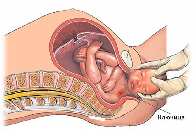
Most often, a fracture of the collarbone in a newborn occurs when an attempt is made to apply gentle traction by the head, or to extract the child by the chest at a time when the shoulders have not yet been born.
Clavicle injury occurs more often in children with a wide shoulder girdle and heavy weight. A large fetus can appear due to a hereditary predisposition in women with gestational diabetes mellitus.
The most common diagnosis is a fracture of the right clavicle. This is due to the peculiarities of the position of the fetus during childbirth - it takes the first position, in which the right shoulder is born first.
A rare cause of fracture is osteogenesis imperfecta. Pathology can lead to injuries to other parts of the body, for example, the risk of injury to the humerus increases, especially in children in the breech position. Osteogenesis imperfecta is a genetic abnormality, so throughout a child’s life, frequent bone fractures and spinal curvature are possible.
Causes of injury
The shoulder girdle is the widest part of the body in a newborn baby, and due to anatomical factors, it is also the most inactive, so damage occurs during childbirth. Dislocation can also occur at a time when the collarbone is subjected to mechanical stress: pressure, sudden displacement, stretching. When leaving the birth canal, the obstetrician can help the newborn by pulling his hand. Another cause of injury is underdevelopment of joint tissue.
Provoking factors that increase the risk of injury in infants:
- Rapid labor - due to rapid delivery, the fetus does not have time to occupy the necessary anatomical position, which helps to come out unhindered.
- Incorrect actions by doctors to use instrumental or manual methods to turn a child moving through the birth canal.
- A woman has a narrow pelvis.
- The large size of the fetus does not correspond to the female pelvis and significantly complicates its passage.
- Presentation of the baby. The correct position of the fetus is head down. During the delivery process, the cranial bones bear the main load, which facilitates the easy advancement of the child.
- Fragility and underdevelopment of joint tissue.
In most cases, dislocation is caused by doctors’ mistakes and incorrect actions of women in labor during labor.
How does a clavicle fracture appear?
Symptoms of a fracture are most often noticeable immediately. In the first days, the movements of a baby's limbs are chaotic, they do not obey consciousness. Already in the first day it will be noticeable that the trajectory of one hand is different from the second, its swings are less active. It is urgent to show the child to the doctor if his arm does not move.
After a day, redness and swelling become noticeable at the site of injury, and a hematoma may appear. During swaddling, the baby screams loudly. A sharp cry appears when pressing on the collarbone, and at this time a crunch is felt under the fingers. Due to the unpleasant sensations, the newborn's sleep is disturbed and he has trouble latching on to the breast.
First aid for newborns with a broken collarbone
A damaged collarbone in a newborn must be immobilized during childbirth. This is done using a Deso bandage. The arm in a half-bent position is bandaged to the baby’s body. If swelling or hematoma is found at the fracture site, the doctor prescribes vitamin K injections, which are administered intravenously. Traumeel S with an analgesic, anti-inflammatory and regenerative effect or another ointment as prescribed by a doctor is applied to the fracture site. The gel is rubbed all over the handle to obtain a rapid therapeutic effect.
During the entire time that the baby’s arm is in a bandage, it is recommended to place it only on the healthy side. In an infant, the clavicle fracture heals completely after three weeks. And if everything is done correctly, no complications, as a rule, are observed.
Possible complications
To avoid complications, it is necessary to start treatment as early as possible. If the parents did not notice the injury in time and the child was not examined by a neonatologist, after a few days the bone will begin to heal on its own. In this case, callus formation occurs. The consequence of improper bone fusion is the formation of a false joint.
The fracture affects the child’s adaptation period. After childbirth, physiological weight loss occurs, but after an injury, children lose body weight more rapidly, so it is difficult for them to recover and begin to gain weight.

Treatment of birth trauma is comprehensive
The consequences may be associated with a more severe injury or a combination of several injuries. Damage to the brachial plexus is dangerous, which can occur during childbirth in the breech presentation if the fetus is removed by the pelvis. The pathology manifests itself in the form of swelling, hemorrhage and damage to the nerve roots in the brachial plexus. Neurological signs depend on the extent of damage and may include the following:
- Erb's palsy - occurs when the roots of the 5th and 6th spinal nerves leading to the arm are damaged;
- Klumpke's palsy - the lower part of the plexus is damaged;
- total paralysis of a limb - damage to the entire plexus, it is noticeable that the arm does not move after birth;
- phrenic nerve palsy – when the processes of the 3rd and 4th spinal nerves are involved in the pathological process.
If a newborn is diagnosed with birth plexitis or paralysis of the arms, this pathology is not associated with damage to the clavicle or brachial plexus. Spinal injuries lead to paralysis.
Diagnostic methods
The doctor may suspect a fracture already during the initial examination of the newborn in the delivery room. It takes into account how the birth took place. A thorough examination of the shoulder girdle is required in the following cases:
- rapid labor;
- application of obstetric forceps or other techniques;
- difficult birth of shoulders;
- heavy weight of the newborn.
During the examination, the doctor may detect crepitus in the collarbone area. Swelling and hematoma appear no earlier than the next day.
The Moro reflex helps check the combined movement of the limbs. This is spinal motor automatism, which is normally present in infants up to 4–5 months, and then begins to fade away. To check it, the child is placed on his back on a changing table. The doctor sharply hits the table surface with his palms 15 cm from the head. Another way to trigger the reflex is to raise your straightened legs and pelvis above the bed. Manifestations of automaticity go through 2 phases:
- the child moves his arms to the sides and unclenches his fists;
- after a few seconds the limbs return to their original position.
A complete absence of the reflex is observed with skull injuries. If a child moves his arms slightly away, this indicates hypertonicity. When the central nervous system is damaged, the reflex does not occur immediately, but with a slight delay. A clavicle fracture is characterized by the absence of automatism on the affected side.
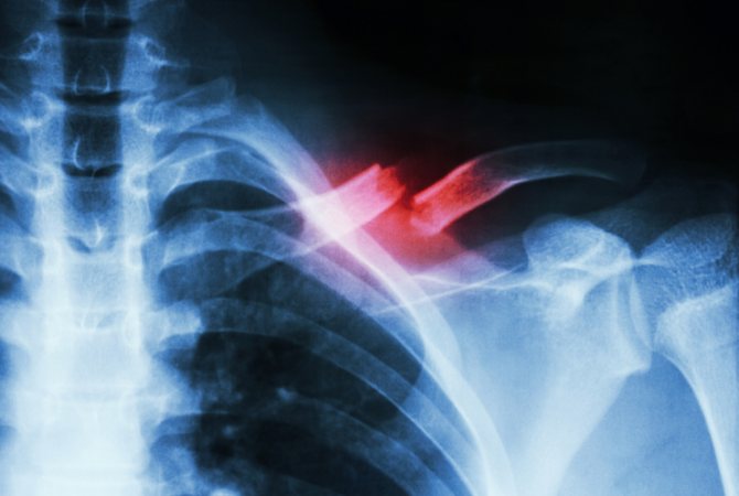
A fracture can be accurately diagnosed using an x-ray. Most often, the injury occurs in the middle third of the collarbone.
Differential diagnosis is carried out with damage to the brachial plexus, injury to the radial nerve, which can occur with a fracture of the shoulder. The latter is characterized by weak extension of the hand, and the thumb hangs down.
During childbirth, the collarbone is more likely to break when the fetus is breech.
Treatment rules
For the success of treatment, it is important to start it as early as possible. In children, reparative processes proceed much faster than in adults. Therefore, after a fracture, a callus may already form on days 3–4, fixing the bone in the wrong position.
After confirming the diagnosis, the doctor places a special cushion in the armpit and fixes the arm in a special position. It is immobilized with a bandage, which is not recommended to be removed. If the child was able to remove the injured arm, it is necessary to consult a doctor again for proper fixation.
In order for the fracture to heal well, the following rules must be followed:
- do not place the child on the injured side;
- During feeding, the affected side should not press against the mother;
- do not remove the bandage for 10–14 days;
- monitor the condition of your fingers.
Blueness and swelling of the fingers in a newborn are associated with impaired blood flow due to compression of blood vessels by a too tight bandage or displacement of the cushion. When this symptom appears, it is necessary to immediately loosen the bandage. Possible consequences are limb ischemia and additional nerve damage.
The course of treatment includes vitamin K for children with extensive hematoma. After the bone heals, a period of rehabilitation is necessary. The child is prescribed massage, exercise therapy, magnetic therapy and electrophoresis. In order for a newborn to gain weight well, the mother’s nutrition must be complete, breastfeeding should be done on demand, but at least every 2 hours.
Damage to skeletal bones during childbirth is often associated with incorrect provision of benefits to children in the wrong position. To prevent these complications, the doctor may decide on the need for episiotomy or perineotomy. An incision in the perineum will cause additional problems for the mother, but will avoid serious injury to the child.
Fracture of the collarbone during childbirth in a newborn: symptoms, treatment and rehabilitation
According to statistics, a clavicle fracture in newborns during childbirth is a fairly common injury.
Usually, an obstetrician-gynecologist makes a diagnosis immediately after birth during an examination of the baby. But in some cases, the injury is noticed after 2-3 days, when hematoma and swelling appear.
Causes of clavicle fracture
The clavicle is a paired tubular bone, which is attached to the chest on one side and to the scapula on the other. A fracture of the right clavicle bone is more often diagnosed, this is due to the physiological position of the child during childbirth.
The main causes of clavicle injury:
- Breech presentation of the fetus. If the position is incorrect, then his clavicle and other bones are severely compressed. The risk of injury increases, especially with rapid labor;
- Narrow birth canal. Discrepancy between the width of the birth canal and the size of the fetus;
- Trauma due to manual or instrumental delivery;
- Imperfect bone formation is a hereditary pathology that increases bone fragility.
Symptoms of a clavicle fracture in a newborn
The newborn is examined by a neonatologist who diagnoses the injury based on the following signs:
- Painful reaction to palpation of the collarbone;
- The supraclavicular region is smoothed;
- After some time, the soft tissues in the collarbone area turn red and swell;
- During palpation, bone fragments rub against each other and crunch;
- With a displaced fracture, the limb moves with difficulty.
The child’s appetite also decreases and he skips feedings. As a result, weight gain is difficult. The immune system is weakened, and there is a risk of infection entering the fracture site.
To confirm the injury, an x-ray examination must be performed.
If the collarbone is broken, but the doctor did not detect the injury after childbirth, then after 7 days a bone callus may appear. Parents should immediately consult a doctor; the pediatric surgeon will order an x-ray to check the correctness of the fusion.
Treatment methods
Fractures in infants heal much faster than in adults; a cast and surgery are only necessary in rare cases. First of all, when a newborn has a fractured collarbone, it is necessary to immobilize the damaged limb.
To limit movements, an elastic and soft Deso bandage is used.
A cotton roll is inserted into the axillary area, the limb is bandaged to the body, but the bandage should not be tight: this can interfere with blood circulation and the child’s fingers will turn blue.
The duration of fracture healing is 7 days. The damaged limb must be fixed for another 30 days in order for its functionality to be fully restored.
If a baby develops a cephalohematoma (extensive hemorrhage with a bulge) in the area of the fracture, then additional treatment is necessary. For this purpose, a solution for injection, Kanavit (vitamin K), is prescribed, which is administered intravenously. The course of treatment is 3 days.
Also, for a hematoma, the doctor may prescribe Traumeel S ointment based on herbal and mineral components. The drug relieves pain, stops bleeding, relieves swelling, and eliminates the inflammatory process. The drug is safe for infants; not only the broken clavicle bone, but also the trapezius muscle can be treated.
For 14 days, the little patient should sleep only on his healthy side or back; during feeding, also turn him over to his healthy side. This is a treatment option for a common fracture or crack in the collarbone.
If the broken clavicle bone has moved, then under no circumstances try to put it back in place yourself . This can damage muscles, important nerves or blood vessels, causing bleeding or muscle paralysis. Go to the hospital where a pediatric traumatologist will provide treatment.
If the displacement is slight, a Deso bandage or Delba rings are applied. And in case of severe displacement, a double Kramer splint is applied. In particularly severe cases, surgery will be required.
In case of a comminuted fracture, parts of the clavicle are fixed with a special splint or plate. Such a fracture heals in 30 days or more.
It is strictly forbidden to self-medicate; if you find that your newborn has a broken collarbone, consult a doctor!
It is necessary to visit a doctor for an examination, diagnosis and proper treatment. In most cases, treatment has a favorable outcome, as the bones in infants heal quickly.
Rehabilitation period
In order for the child to recover faster, it is necessary to restore nutrition to the damaged area and strengthen the clavicle bone.
Post-traumatic rehabilitation includes the following procedures:
- Magnetotherapy – the effect of a multi-frequency magnetic field on the damaged area;
- Physical therapy also promotes recovery. The mother performs simple exercises: it is necessary to move the damaged limb away from the body, and then return it to its place;
- Massage promotes rapid healing, but should only be performed by a specialist. After the bone has fused, the mother can massage the shoulder on her own, this improves blood circulation;
- Electrophoresis is a physiotherapeutic method in which current and a special drug are applied to the body in order to restore it after injury;
- Proper nutrition is the key to a quick recovery. The best option for a baby is mother's milk, otherwise you need to eat foods that contain a lot of calcium, phosphorus, magnesium and silicon.
Consequences and complications
Competent and timely therapy guarantees rapid bone healing. In some cases, when the newborn's immune system is weakened, an infection can spread to the fracture site. A false joint may also form if the bone was not properly fixed or the fracture was severely displaced.
For severe pain, the doctor prescribes analgesic drugs that are administered intravenously, but this is only possible in inpatient treatment conditions.
Pain syndrome occurs if the brachial plexus or neurovascular bundle is damaged . But such complications arise in isolated cases.
Thus, a clavicle fracture is a common birth injury that does not pose any particular danger. An experienced doctor will identify the disease immediately after birth and prescribe proper treatment. Do not self-medicate, monitor the child’s condition and, if in doubt, go to the hospital.
Victor Systemov – expert of the 1Travmpunkt website
Source: https://1travmpunkt.com/perelom/raznie/klyuchicy-u-novorozhdennogo.html
User comments
Hello. We have a collarbone fracture. 1.5 years. It also grew together incorrectly. One bone sticks up, the other down. Shorter with offset. How did everything work out for you in the end? What the control scan showed. Thanks in advance. I'm very worried about my daughter.
It grew together perfectly) but what bothered me (the bump on the bone) was a bone callus, which resolved within a few months.
Thank you, Anna. You reassured me. We were told to take a test in a year.
The kids are getting along just fine. And the bones break like a green branch. So everything will be fine.
What happened to your collarbone in the end, otherwise we have the same problem now.
Do not worry. The broken collarbone should heal like that. I myself broke my collarbone as a child. With offset. (I understand that you also had a displacement). And I wasn’t so small, I was already a healthy aunty horse, in the 9th grade. I also didn’t get a plaster cast, but rings. And it worked out the same way for me. They didn't break anything. The doctor said it was normal. Children often grow together like this. It’s just that later the bone grows and this place will not be visible. Over time everything will be fine, everything will get better and it will be normal, I assure you! No one will break or edit. The child is small, grows quickly, the bones are not ossified like in adults. Over time, they will level out on their own and grow.
This is what happened to me. And now, many years later, I don’t really remember which collarbone I broke, right or left, and they both feel the same!
Do not panic! Everything grew together fine for your baby. Just take care of the handle for a while so that he does not lift anything heavy, and preferably so that he does not fall on it. Just keep your eyes peeled.
I repeat once again: they won’t break it, everything will be fine over time and you and the child will forget this collarbone like a bad dream.
In order to correctly assess the patient’s condition, make an accurate diagnosis and prescribe appropriate treatment, it is necessary to accurately determine the type of clavicle fracture, the presence or absence of displacement and associated complications. All this significantly affects the recovery time and healing of the broken bone in general.
Classification of clavicle fractures
Clavicle fractures in children are classified based on seven factors:
- by time of occurrence (congenital or acquired);
- due to occurrence (trauma or pathology);
- by skin integrity (open or closed);
- at the location of the fracture (intra-articular, periarticular, extra-articular);
- by type of damage to the periosteum (without damage or with rupture);
- in the direction of the fracture (transverse, longitudinal, helical, spiral, annular);
- by the presence of displacement or its absence.
Complications often accompany a clavicle fracture. These are damage to soft tissues, blood vessels, subclavian veins or arteries, and brachial plexus trunks.
Features of trauma in children at different ages
Clavicle fractures in preschool children are fractures or subperiosteal fractures. In the photographs they look like a “twig” - the layer of periosteum holds the bone fragments in place, the fragments are not displaced relative to each other, the bone externally retains its integrity, but is broken and bent. In schoolchildren aged 7 years and older, the layer of periosteum is thin, so the previous retaining effect is not created.
With age, the severity of fractures increases, and complete fractures occur more often. The degree of displacement of the fragments varies greatly, and this can significantly injure surrounding tissue, as can be seen in the picture.
Types of fracture
Based on the location of the bone fragments, the following types of injury are distinguished:
- Displaced clavicle fracture. The bone fragments are displaced relative to each other. Displacement occurs with fractures such as:
- complete - the wreckage is scattered.
- incomplete - the integrity of the bones is largely preserved.
The collarbone is broken without displacement. Such injuries heal much better.
But most often closed fractures occur. With such injuries, the skin remains intact.
According to the fault line, a broken collarbone is divided into the following types:
- T-shaped.
- Transverse.
- Oblique.
- Helical.
- S-shaped.
This division of fractures is of great importance, since the choice of effective treatment, without serious consequences, depends on the type of injury.
Important! A clavicle fracture in children occurs due to the fact that the bone tissue is soft, but they are able to quickly recover after birth trauma.
Diagnosis of fractures with and without displacement
Diagnostic methods used in combination help to diagnose a clavicle fracture, identify displacement and other injuries. There are situations when an experienced neonatologist determines a clavicle fracture in a newborn based on the existing clinical manifestations:
A pediatric neurosurgeon, thoracic and vascular surgeons are additionally involved in the diagnosis. A wide range of diagnostic methods are used to determine in more detail the range of injuries that caused the fracture and determine the correct treatment tactics.
What is the clavicle
To understand why a clavicle fracture occurs in newborns, you must first become familiar with the structure of the children's skeleton. The collarbone is an important component of it. This S-shaped bone connects the sternum to the shoulder blades. This organ is a pair; each person has a right and a left collarbone.
Dice Features:
- Miniature sizes;
- When raising the shoulder girdle, the movement of the bone resembles turning a key in a keyhole, which is how it got its name;
- The fragility of the bone is compensated by its location, the likelihood of injury in adulthood is minimal.
According to the classification, the clavicle belongs to the long bones of the skeleton, but it is not hollow, but spongy, like short bones. This is another of its features. If the integrity of the clavicle is violated, there is a high probability of destructive changes in the structure of the upper limbs. For this reason, the fact that a newborn has a fracture cannot be ignored.
In children, bones knit together at record speed. This is both an advantage and a noticeable disadvantage in the process of treating birth injuries. If a violation of the integrity of the clavicle is not detected in a timely manner, there is a possibility that it has already fused incorrectly. Then the child will have to have a serious operation.
Fracture treatment
Such a fracture is treated by a pediatric traumatologist. Only with qualified help can you count on successful restoration of the functions of the shoulder girdle. Treatment by a specialist will ensure that there are no additional injuries in most cases. Independent attempts to straighten the bones are fraught with complications.
There is no doubt that if a fracture is suspected, it is necessary to take the child to the emergency room as quickly as possible, if possible, or call an ambulance. Before the doctors arrive, you can give the child a painkiller (Analgin) and first fix the injured arm with a scarf and then bandage it to the body. To reduce the risk of swelling, an ice compress is applied to the fracture site - a container of ice wrapped in a cloth to prevent hypothermia.
Recovery of babies under 1 year usually occurs in 12-20 days. In a medical institution, the child’s arm is bandaged to the body for seven days, while a small cotton swab (Dezo bandage) is placed under the armpit. It is imperative to monitor the color of the injured hand - at the slightest appearance of cyanosis, the bandage is loosened. Put the baby to bed and breastfeed only on the healthy side. Young children are given a Deso bandage in the same way as babies from 0 to 12 months. In children 4-5 years of age and older, it is not always possible to fix the arm with such a bandage, so other methods are used:
- A pair of rings are placed on the hands up to the level of the armpits and pulled together at the back. This slightly stretches the muscles, but does not guarantee fixation of the bone fragments. This way you can fix the fracture for up to two months.
- A softer method of fixation is a figure-eight bandage, the ends of which are brought out on the back.
- A plaster cast is the most rigid method of fixation. Before applying it, it is necessary to numb the injured area. With its help, the fracture is fixed in the correct position for up to 21 days. To decide whether to remove the rigid cast, a repeat x-ray is necessary to confirm bone fusion. You can see in the photo how the plaster is applied.
- The most bulky but reliable design is the Kuzminsky tire. With its help, you can create displaced fractures within 2-3 days. Patients are advised to sleep on their back.
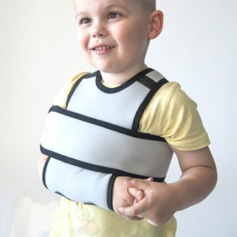
- Lightweight options for fixing fractures have now been invented and are widely used - bandages and corsets made of lightweight, synthetic, hypoallergenic materials. They provide fairly rigid fixation of the injured limb and have a long service life. Such orthopedic devices can be found and sized in special stores.
For surgery for a clavicle fracture, the child must have serious reasons:
- open fracture;
- impossibility of combining debris;
- fracture with many fragments;
- damage to blood vessels, nerves or lungs.
The operation is performed on young children under general anesthesia. For older children, local anesthesia with Novocaine or Lidocaine is sufficient.
- special plates of various shapes;
- LCP plate;
- pins and screws for insertion into the collarbone.
Osteosynthesis is completed with a Deso bandage. Surgery is the most traumatic method. However, it has a number of advantages:
Complications of osteosynthesis are very rare:
- Non-union of the clavicle if the fixator is chosen incorrectly or the fracture is comminuted.
- Infection (osteomyelitis) that occurs when the rules of asepsis are violated (we recommend reading: symptoms of osteomyelitis in children). An antibiotic administered intravenously half an hour before surgery helps prevent infection. The same drug is then continued orally for 7–10 days.
The retainers are removed through another operation. It is carried out no earlier than in a year.
Treatment methods for fractures and dislocations
Treatment of a fracture in a newborn depends on the type of injury, when it was discovered, and possible complications. Therapy consists of immobilizing the arm and prescribing medications that will help relieve swelling and restore tissue. In the case of newborns, fractures heal much faster than in adults.
A clavicle fracture during birth can be of two types - with and without displacement. In most cases, there is no displacement, and this type of fracture heals quickly. After identifying the injury and examination, a tight bandage is applied to the injured area, while the mobility of the affected limb is limited so that the baby does not harm himself with unconscious movements.
Dislocation is treated in the same way. The exit of the bone from the joint can be easily detected, and a pediatric orthopedist or traumatologist can handle the reduction. The specialist applies a bandage, which, depending on the complexity of the injury, parents can change at home themselves. Bandages must be applied carefully, without making the loops too tight, so as not to disrupt blood circulation in the injured limb. Traumeel C ointment is used to reduce inflammation.
In some cases, the attending physician uses analgesics: they are needed if the pain is severe and causes loss of appetite. Pain syndrome develops only in rare situations when the neurovascular bundle is affected. Most often, it does not cause severe discomfort and does not require medication.
If there is displacement of the fracture, more complex types of bandages are applied to fix the fragments. Surgery may be necessary only in the most difficult cases.
The rehabilitation period after a fracture
Restoration measures are as follows:
- Magnet. Exposure to a multi-frequency magnetic field at the site of injury.
- Exercise therapy. These are light exercises with the arm, tilting it to the sides, careful bending.
- Massage. A specially selected set of manipulations, which is first performed by a specialist, and then you can do it yourself.
- Electrophoresis. Traditionally used to accelerate the healing of fractures and stimulate blood flow.
- relieve pain;
- reduce inflammation;
- activate healing.
After three weeks, it is recommended to do exercises to warm up the clavicle joint: shoulder movements within the limits allowed by the bandage, then add movements to the shoulder girdle and shoulders. Exercises are needed so that after removing the bandage, the joints are ready for vigorous, painless movement.
The recovery period depends on the patient’s age, the degree of complexity of the fracture and can last up to two months. During treatment, you must strictly follow the instructions of the traumatologist. This is the only guarantee that the bone will heal correctly.
A clavicle fracture in children is a common injury that many children receive during active play or from a fall, for example, while riding a bicycle or roller skating. Often parents cannot correctly recognize the child’s condition, and a broken collarbone is confused with an ordinary bruise and does not pay special attention to it.
But this position can lead to serious consequences, because it is important to take measures on time, therefore, if the baby falls, hits his shoulder, and a bruise appears in the collarbone area, you need to consult a traumatologist.
If measures are taken on time, then, as a rule, there is no further discomfort from the fracture, since bones in children heal easily and quickly. But it is important to make a correct diagnosis, because in case of an incomplete fracture this can only be done with the help of an X-ray examination.
Therapeutic measures
The fact that the bone has left its anatomical position can be easily detected by a doctor during a visual examination of the child. Conservative therapy consists of a reduction procedure. At the Center for Sports Traumatology and Rehabilitation Medicine, after repositioning, a bandage is applied, which is necessary to fix the collarbone and prevent re-dislocation. The bandage should not be applied too tightly so as not to disrupt the blood circulation of the upper limb. If the case is not severe, parents can change the fixing bandages themselves.
To eliminate edema as quickly as possible, local spectrum drugs – ointments and gels – are used. Rarely, if a child is experiencing severe pain, painkillers are prescribed. But, as a rule, they are not needed. Severe pain syndrome occurs only when nerve endings are affected. If they are still affected, analgesics are administered to the infant intravenously, exclusively in a hospital setting.
During the period of fixation of the collarbone and for some time after removing the bandage, until complete recovery is complete, parents need to monitor how the child sleeps. You should not lie down on your injured side. You are allowed to sleep either on your back or on your healthy side. Since it is quite difficult to constantly monitor a baby’s sleep, it is recommended to immediately lay him in the desired position, and place a towel bolster or pillow on the side that will not allow him to roll over in his sleep.
Types of fixing bandages
Based on the severity of the traumatic injury, the doctor chooses one of the methods of applying a bandage. In infants, fixation of the injured limb is of great importance because their arms are constantly in motion. And this can lead to displacement of the end of the clavicle and provoke complications.
Fixation methods:
- The Volkovich method is the application of a thick cotton-gauze roll, which is secured on top with an adhesive plaster. The shoulder on the injured side is slightly retracted and outward. When completing the bandaging, place a cotton swab in the armpit. The hand is lowered down and tied using the scarf method.
- Plaster immobilization is the most commonly used method, which allows you to completely immobilize a limb.
- Dezo method - the shoulder is pulled back, a cotton-gauze bandage is applied, and it is fixed with a plaster. The bandage covers the shoulder girdle and the lower third of the shoulder. To achieve high-quality immobilization of the hand, a second strip of adhesive tape is additionally applied, which is positioned perpendicular to the first.
Fixing bandages make it possible to begin rehabilitation a few days after their application. The first thing you need to do is passive exercises to keep your muscles toned. Parents should carefully bend and straighten the child’s fingers and make circular movements with their hands.
Surgical intervention
If the injury is not detected in a timely manner, after 1-2 weeks it will be considered old. Stale lesions are mostly treated surgically. The operation is also performed if conservative methods - reduction with further rehabilitation - have given only short-term results, and the dislocation occurs again.
There are many techniques for surgical treatment of clavicle dislocation. At the Center for Sports Traumatology and Rehabilitation Medicine, the operating method is selected individually for each clinical case. The safest method is minimally invasive, arthroscopic intervention.
This type of surgical intervention does not require incisions of the skin and soft tissues, and therefore there is practically no risk of a number of complications characteristic of open operations. The rehabilitation period is also significantly reduced, which is especially important in the treatment of young children.
Today, the most effective and safest method is ligament plastic surgery. This method involves the surgeon creating artificial tissues that are implanted in place of damaged tissues that cannot be restored.
Physiotherapy
The rate of restoration of joint tissue in an infant is high. But since it is very difficult to limit a baby’s movements, the risks of re-injury are high. Physiotherapeutic techniques are used to speed up recovery:
- Magnetotherapy is the effect of a magnetic field of different frequencies on the damaged bone.
- Electrophoresis – promotes better penetration of topical drugs.
- Massage improves blood circulation, which in turn delivers the necessary amount of nutrients and oxygen to the joint elements.
Features of fractures and their classification
This is why a closed fracture of the clavicle is often confused with an ordinary bruise, because the main symptoms are not pronounced, the child does not feel severe pain, but only complains of discomfort when moving his arm.
A fracture can be classified according to many factors, for example, according to the condition of the skin, they distinguish:
- closed type, when the skin is not damaged, but there is swelling and bruising in the collarbone area;
- open type with damage to the skin, sometimes with fragments protruding from the wound - in this case the child will need urgent surgery; It is important to see a doctor quickly to avoid possible infection.
Based on the degree of separation of bone fragments, two types are distinguished:
- without displacement of the broken bone;
- with displacement - such a fracture can be either incomplete, when the integrity of the bone is largely preserved, or complete, if the connection between the resulting fragments is completely broken.
According to the bone fracture line, a clavicle fracture is divided into:
- transverse;
- longitudinal;
- helical;
- splintered;
- oblique.
Diagnostics
First of all, when diagnosing a clavicle fracture, symptoms are taken into account. With a complete fracture, which is considered a classic form of injury, there is usually a tear of the periosteum. The main sign of a fracture in this case will be a feeling of pain in the injured area, as well as the appearance of swelling, which is caused by the divergence of the damaged bone.
An injured shoulder always looks shorter than a healthy one, since the impairment is located between the neck and shoulder joint. The shoulder blade droops slightly, and its damaged edge begins to protrude much more sharply than usual. This occurs because the broken collarbone can no longer hold the shoulder blade in its natural position. The arm also shifts, it turns inward and protrudes a little forward, and normal movements cause a feeling of pain, especially if the child lifts it up from the side.
At first signs, such a fracture looks like a simple bruise, but if no action is taken, then in just a week a dense tumor (callus) will appear at the site of the injury. In this case, you must urgently contact a specialist. An accurate diagnosis, as well as identifying the features of a fracture, is possible only after an x-ray.
Treatment
A patient with a clavicle fracture, regardless of his age, must be given first aid, but in no case should you try to reconcile the broken bone yourself. It is important to remember that when trying to align a fracture, nerves, muscle tissue or blood vessels can get caught between the bone fragments, and this can cause a lot of serious complications, including paralysis.
First aid measures should be:
- Give a painkiller.
- Bend the arm on the side of the injury at the elbow and fix it to the body, providing the injured area with the necessary rest.
- Take the child to the doctor. This could be a surgeon in a regular clinic or a traumatologist at the nearest emergency room.
Treatment of clavicle fractures can be carried out in several ways, depending on the characteristics of the injury. In children with a clavicle fracture, a fixing bandage is usually applied, and treatment has a certain sequence:
- First, the injured area is numbed.
- Then the fragments of the clavicle are combined into the correct position, while placing a thick cotton-gauze roller in the armpit of the small patient.
- The shoulder must be immobilized, since bone fragments can easily be dislodged, and this can lead to malunion and complications in the future. To fix the damaged area, bandages are usually used: Deso or figure-of-eight.
Plaster casting is another way to treat a clavicle fracture, and in 95% of cases it heals completely, but it is important to remember that a plaster cast alone will not eliminate the existing displacement of the fragments, so before installing it, the damaged bone must be aligned in the correct position.
If the injury is not corrected, then after removal of the plaster, one may find deformation of the shoulder joint, its shortening, due to the fact that the normal length of the shoulder girdle has not been restored.
If at the same time there is a thickening of the clavicle formed at the site of the fracture, this indicates that the existing fragments have grown together. In the future, the callus may decrease, but the deformation of the clavicle due to improper fusion will not be eliminated.
If it was not possible to manually eliminate the violation when the fragments of the clavicle were displaced, it is recommended to perform osteosynthesis - a special operation during which all bone fragments are folded into the desired natural position.
Treatment and first aid for injury
The first measure that is necessary when a clavicle fracture is detected in a newborn is immobilization.
For this, a Deso bandage , as it is elastic and soft. It is used to secure the child's hand to his body. In the presence of extensive hemorrhage with a bulge, which is called a cephalohematoma, intravenous vitamin K is additionally prescribed.
Traumeel S ointment is usually chosen as a remedy that has an analgesic effect , which can also be successfully used as a remedy against edema. In addition, it can slightly increase regeneration. It is important to lubricate the entire trapezius muscle, not just the broken collarbone.
For two weeks, the newborn should not lie on the side on which the bone was broken. Treatment of a clavicle fracture in newborns after discharge continues at home. After 20 days, the bone tissue will completely heal and this injury will not affect the child’s health in any way.
Timely first aid is half the success in treatment
Usually, a fracture of the collarbone in children is diagnosed almost immediately after birth, but sometimes it happens that such damage occurs in infants after discharge from the maternity hospital.
In this case, it is important to provide first aid on time, because a lot depends on its correctness and timeliness. There are two ways to fix the hand in the presence of a fracture of this kind:
- You should fold your child’s arms on his chest, first bending them at the elbow, and then place them behind his head. Next, a stick should be placed behind the back, which should be held in the elbow bends. But since not every child can lie like this for a long time, it is better to use the second method.
- It is necessary to immobilize the baby's limb, and a bandage that is not too tight is suitable for this purpose. The hand should be suspended on the scarf at a right angle.
If a baby shows pallor, cold perspiration, and a pulse rate, we can say that he is developing all the signs of vascular insufficiency.
In this case, you can give the child a little sniff of ammonia.
Afterwards, the baby should be taken to the hospital immediately.
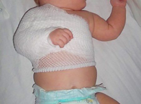
The photo shows a Deso bandage used for a clavicle fracture in children
Pain relief can be achieved through the use of special ointments, as well as through injections.
Solutions are injected directly next to the injury site so that they act faster.
Since the arm should be immobilized for the entire duration of treatment, a special type of bandage is selected that is most suitable for children. These include the following:
- Delbe rings. Also suitable for fixing displaced fractures. It consists of two rings placed on the patient’s shoulders and connected by a bandage. They tighten the shoulder blades, immobilizing the injured arm.
- Deso bandage. It is performed by fixing the arm to the chest using bandages surrounding the chest and shoulder.
- 8-shaped bandage of fixing type.
- Crutch-plaster cast.
Features of lifestyle and rehabilitation
The bandage applied by the doctor cannot be removed independently during the entire treatment period. The baby's physical activity should be limited. An exception is physical therapy exercises, which are necessary for proper restoration of muscle tone and mobility of the damaged area.
If the fracture was incomplete, the child will have to wear a fixing bandage for up to 3 weeks, but if there are displacements, the healing period usually increases to a month. Full functionality of a hand with a damaged collarbone will be restored only after 2-3 months of rehabilitation.
In the first days after applying a therapeutic bandage, the child should begin to move his fingers and elbow joint, but with caution, avoiding pain. After removing the fixator, the doctor will prescribe special procedures in the physiotherapy room, the purpose of which is not only to eliminate pain and discomfort, but also to speed up the healing process of the fracture, relieve the inflammatory process and improve the condition of the little patient.
For rehabilitation, the child is prescribed the following procedures:
- ultrasound therapy;
- magnetic therapy;
- laser therapy;
- UHF using special anti-inflammatory ointments.
After a three-week course of treatment, the child will be able to move the shoulder within the limits of the applied bandage. At this time, therapeutic exercises are expanded, and in addition to moving the fingers and elbow joint, it is allowed to shrug the shoulders, pull them back and do other exercises to warm up the joint and restore its mobility.
After the doctor removes the applied bandage, the joints of the arm and injured shoulder will be relatively functional, and full mobility, subject to all the instructions of the specialists, will be restored very quickly. Often, at the end of rehabilitation, the child is prescribed a special massage and lotion or mini-bath with sea salt.
Treatment and healing time for a crack in the collarbone in an adult and a child
Treatment of clavicle fractures can occur conservatively or surgically. The duration of the rehabilitation period depends on the quality of the developed treatment regimen, as well as the individual characteristics of the patient’s body.
Conservative method of therapy
After anesthetizing the damaged area, the doctor performs a closed reposition of the fragments. To do this, the patient sits down on a chair and tilts his head in the direction opposite to the injury, since this allows the bone fragment to be brought closer to its anatomical location, as well as to relax the muscles. The duration of the procedure is 10-15 seconds.
If the doctor fails to compare the fragments after three attempts, the patient is prescribed surgery.
Approximate duration of bone fusion is 1.5 months
After the fragments have returned to their anatomical position, the patient is given a cast on the damaged collarbone, which must be worn until the end of healing. As a rule, the chest, shoulder girdle and forearm also fall under the plaster cast. The approximate duration of bone fusion is 1.5 months, during which time X-rays are regularly taken to monitor dynamics. There is cause for concern in the following cases:
- tissue swelling has increased;
- the skin above the lesions becomes hot to the touch;
- the skin at the site of injury is inflamed and red for more than five hours in a row.
Alarming signs indicate the development of an inflammatory process and require adjustments to the therapeutic course.
Surgical method of treatment
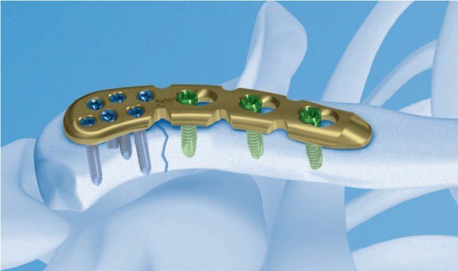
Osteosynthesis of the clavicle with a plate
Surgical intervention in most cases is required for open types of injury, if the examination results showed a large number of fragments, there is a possibility of damaging soft tissue with fragments, or the previously broken bone has not healed properly.
Before proceeding with the operation, the patient is given general anesthesia. Osteosynthesis can be carried out inside the bone, on it or outside using a special fixation apparatus.
Drainage is brought to the surface from the wound, which is removed several days after the operation, provided that healing is going well. For the first few days, the site of injury must be immobilized, and then a permanent cast must be applied. After two weeks, the patient’s sutures are removed, and a month later a control X-ray examination is performed and, if there are no deviations from the norm, the plaster cast is removed.
Features of a clavicle fracture in newborns
Childbirth is a complex physiological process, and its outcome can never be predicted 100%. Often the birth of a baby is accompanied by various injuries, among which the most common is a fracture of the collarbone. This can be due to various reasons, for example, a very large fetus or a narrow pelvis of the expectant mother, rapid labor when the newborn does not have time to make all the necessary turns.
Sometimes it is not possible to make a correct diagnosis for a newborn right away, since in most cases the baby’s skin has a changed color, even bluish, if the birth was especially difficult. For this reason, doctors may not notice bruising or swelling at the fracture site for some time. Having discovered such symptoms, the newborn is given an x-ray and measures are taken to eliminate the injury and fix the handle.
In this case, a soft fixing Deso bandage is used as treatment, and the baby’s arm is bandaged to the body. In some cases, the doctor may prescribe vitamin K injections intramuscularly for 3 days, which is necessary if there is a severe hematoma. Pain-relieving ointments are also used in treatment, and they are applied not only to the fracture site, but also to the trapezius muscles.
For two weeks, the newborn should not lie on the injured side; he can only lie on his healthy side or on his back. But it is important to change position frequently, since the baby’s skull bones remain mobile for several months after birth, and if he constantly lies on one side, deformation of the shape of the skull with facial distortion may occur.
In the maternity hospital, the mother is taught how to properly apply a fixing bandage and carry out the necessary procedures, so that after discharge from the maternity hospital she can cope with all the difficulties herself and continue treatment. As a rule, a clavicle fracture in an infant heals within 3 weeks, after which motor activity is quickly restored. In most cases, such damage does not affect the future health of the baby.
Treatment tactics
The first thing you need to start with is to fix your hand using swaddling. Also for this purpose, a Deso bandage can be used, which allows you to fix the handle to the body. If there is a cephalohematoma (hemorrhage under the periosteum) or ecchymosis (extensive bleeding into the skin), vitamin K is indicated for the baby; it is injected into the muscle for three days.
For two weeks, the child should not be placed on the affected side; he should sleep and eat on the healthy side or on his back. After healing of the fracture, the function is completely restored, and the fracture itself does not affect the child’s health in any way.
Treatment of a broken collarbone in a child (techniques in the photo)
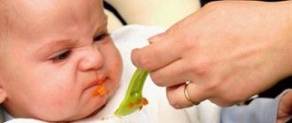
Correct position when eating
Imagine the best day of your life? Surely many will name the day when their child was born. Indeed, the birth of a new life is the most wonderful event for a married couple. For 9 months, the expectant mother and father dream of seeing their child as soon as possible, unaware of the possible danger. Childbirth is an unpredictable process, but this does not mean that you cannot have children. It is important to be prepared for possible complications and provide first aid in a timely manner.