You can assess your health status not only by blood tests. Often, to diagnose pathologies of the body, a nasal swab is taken for eosinophils. This is a group of leukocyte cells that are responsible for the protective functions of the immune system. If the results of a laboratory test show deviations of eosinophilic cells from the norm, then doctors will be able to understand what exactly provokes the disorders by their increased number.
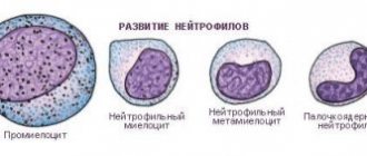
Eosinophils (EOS) are indivisible granulocytes. They, like other blood cells, are formed in the bone marrow. The process of cell formation occurs within 3-4 days. They then migrate into the bloodstream, where they circulate with red cells for about 6-10 hours.
From the vascular network, eosinophilic forms move to different organs. They are mainly localized in the digestive tract, respiratory system, and skin. Life expectancy is on average about two weeks. After the death of some eosinophils, new mature formed elements come to replace them.
Why are eosinophils needed: functionality and normality
- Destroy helminths;
- Absorb foreign bodies.
The level of eosinophil cells depends on the daily production of the hormone cortisol. The norm of leukocytes changes with the patient’s age, but ideally there should not be any of them in the smear at all. If they are present in small quantities, then the following figures are considered acceptable indicators (measured as a percentage, %):
- Children under 13 years old – 0.5-7
- Teenagers and adults – 0.5-5
Single eosinophilic cells in a smear indicate a weak pathological process or the initial stage of some inflammatory process.
Nasal swab for eosinophils - interpretation and norm in children and adults
In a nasal smear in a child and an adult, special attention is paid to the number of desquamated columnar epithelial cells and the number of leukocyte cells. The obtained research information is used in conjunction with the medical history, clinical picture and the results of other diagnostic techniques.
The table shows the values of different types of leukocytes in the analysis of nasal secretions for healthy people.
It should be emphasized that about 10% of the cells in the smear cannot be differentiated due to the lack of clear cell boundaries or a large amount of mucus. The shares of ciliated and flat cells account for 1 and 10%, respectively.
Important: detection of a small number of representatives of the normal microflora of the nasal cavity (usually coccal bacteria) is allowed.
In what cases is a rhinocytogram prescribed?
The collection of mucous material from the nasopharynx is carried out mainly during long-term rhinitis of unknown etiology. Eosinophils from a rhinocytogram make it possible to understand what kind of runny nose the patient has - allergic, infectious or other origin.
An increased number of eosinophilic cells in a nasal smear is observed with allergies. If the diagnosis shows a significant deviation from the norm, then the doctor will be able to prescribe the correct treatment, because the treatment of a viral or bacterial runny nose is significantly different from allergic rhinitis.
Examination of the nasal mucosa makes it possible to see the level of not only eosinophils, but also other blood fractions. A rhinocytogram will show the number of neutrophils, macrophages, and lymphocytes. Also, normally, cocci, yeast fungi and other non-pathogenic microflora are found in the biomaterial in small quantities.
The diagnosis is deciphered after counting all the blood cells and microorganisms in the patient’s mucus. Doctors do not take into account only one of the indicators, but always assess the state of a person’s health according to other parameters. Here's what you can understand from the results of a rhinocytogram:
- Red blood cells - their presence in the mucous biomaterial indicates the high permeability of the vascular network in the nasopharynx (this is characteristic of infectious diseases such as influenza, diphtheria, etc.);
- Lymphocytes – usually present in chronic nasal inflammation;
- Neutrophils - observed in acute infections that cause a prolonged runny nose;
- Eosinophils - speak of allergic rhinitis, the development of polypous tissue in the sinuses.
A rhinocytogram, which did not reveal an increase in eosinophilic bodies or other fractions of leukocytes, allows us to conclude that a prolonged runny nose is caused by vasomotor reactions, addiction to vasoconstrictor drugs, impaired nasal anatomy or hormonal imbalance in the body.
High eosinophils in a nasal smear: causes
Of course, a nasal swab alone cannot confirm any diagnosis. He gives directions to the doctor in which direction to move in order to find out the exact cause of the disorders. But first of all, a rhinocytogram makes it possible to detect allergic rhinitis or pathological processes of another etiology at an early stage.
False-negative results are obtained by patients who, before diagnosis, instilled medications with hormonal components (corticosteroids) into their nose. The same effect occurs when taking antihistamines. Therefore, if you need a correct interpretation of the tests, then you should exclude treatment with such medications several days before the procedure.
Deciphering the rhinocytogram in children: the cellular composition of a nasal smear.
Rhinocytogram decoding in children is carried out under a microscope. The material for the study is a nasal swab (from the mucous membrane). The collection of material is absolutely painless; in addition, the method has no contraindications.
After applying the material to a glass slide and special staining, a laboratory doctor examines the cellular composition of nasal mucus under a microscope. Normally, the main cells when deciphering a child’s rhinocytogram are neutrophils (65-70%), other cells are represented in significantly smaller numbers: eosinophils (0-5%), lymphocytes (0-5%), monocytes (0-1%), basophils and red blood cells are absent. In addition to the main cells, the rhinocytogram contains cell nuclei that cannot be differentiated (on average, 5-10%), and fragments of the epithelium covering the inside of the nasal passages: ciliated cells - 0-1%, flat cells - 0-10%. A moderate amount of bacteria (mainly coccal flora) and mucus is also a completely normal finding when deciphering a rhinocytogram in children.
If children have an allergic rhinitis, how can one understand the nature of the course based on the symptoms?
Often, even before diagnosing mucus from a child’s nose, parents can already guess what exactly is causing the long-term runny nose. In childhood, persistent rhinitis occurs quite often. Moreover, if children are allergic to various irritating particles, then parents have to constantly fight with the provocateurs of catarrhal discharge.
Allergic rhinitis can be seasonal or year-round. The first option is caused by seasonal allergens, such as pollen or insect bites. A year-round runny nose is observed with allergies to household dust, paper mites, food products, etc.
Using the table, parents can see the difference between the symptoms of the two types of allergic rhinitis.
If a child has such symptoms of a prolonged runny nose as in one of the groups described, it means that he is worried about an allergic reaction. Treatment of such a problem is quite complex and requires a lot of effort on the part of both children and parents in order to eliminate contact with the allergen and maintain health at a normal level. The sooner you start therapy, the easier it is to get rid of the ailment and prevent the development of complications.
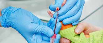
How is biological material collected from the nose?
If someone is afraid of diagnostic procedures, then they can be comforted by the fact that a rhinocytogram is an absolutely painless and very quick manipulation. Even small children tolerate the collection of mucus from the nasopharynx well, although it causes minor discomfort.
To make it less scary and clear how the procedure occurs, we will tell you the step-by-step process of carrying it out by a laboratory assistant:
- The specialist takes a thin and long tube with a cotton wool tip.
- Introduces it carefully to the posterior part of the inferior nasal concha and scrapes off the mucus with a slight movement.
- He takes out the tube and packs it in a special container or a separate sterile tube, in which it is transferred to the research room.
In the laboratory, the resulting sample of biomaterial is stained to count the number of eosinophils and other formed elements. Eosinophilic bodies acquire a pink tint, separating them from other cells and microorganisms.
Taking a smear is not a procedure for which it is necessary to carefully prepare the nasopharynx or the body. Unlike donating blood, where it is important to adhere to a diet, avoid bad habits, physical influences and stressful situations that distort indicators, a rhinocytogram does not require compliance with such rules. But still, recommendations, albeit limited, are given to patients.
- Before manipulation, it is better not to intensively blow out the mucous secretions, so that the real amount of formed elements remains in the analysis.
- Do not instill intranasal drops into the nasal cavity (applies to any means - antibiotics, vasoconstrictors, etc.).
These are all the rules that you need to remember before visiting the diagnostic center. For more reliable results of mucus sampling, it is recommended to repeat the manipulation after a few days.
Many patients are interested in where they can get a nasal swab and what the cost of such a diagnosis is. There are laboratories in various clinics and clinical centers. Almost every medical institution that performs a blood test also collects mucus from the nasopharynx and analyzes the indicators. The biomaterial can be submitted to Invitro or any other laboratory.
But as for the cost, it also depends on the medical institution. If this is a paid diagnostic center, then the price will be acceptable, but not high. And in public clinics this procedure is either completely free or very cheap. Of course, the more seriously the laboratory takes its work, the more reliable the result of the smear analysis will be.
The human body has a unique structure, because each of its systems is responsible for specific functions. Eosinophils, one of the types of leukocytes, also play an important role in the life of the human body. To determine the number of these cells, a blood test is performed or the patient’s sputum is examined, for this a nasal swab is taken.
What are eosinophils?
Eosinophils under a microscope
Eosinophils are non-dividing granulocytes that are formed from a single bone marrow stem cell. The process of formation of eosinophilic cells occurs on average in 3–4 days, after which they are released from the bone marrow and circulate freely in the blood for 6–2 hours. The lifespan of eosinophilic leukocytes varies from 10 to 14 days. From the blood, cells move to the gastrointestinal tract, lungs and skin, where they remain for the entire period of their life.
The presence of eosinophilic leukocytes is very important for the full functioning of the human body, because these cells perform the following functions:
- Have a destructive effect on helminths;
- Absorb foreign particles and cells.
Fluctuations in the level of eosinophils in the blood depend on the daily production of cortisol in plasma.
Normal eosinophil count
To determine the number of cells in the human body, specialists perform a blood test. Sometimes, to study the quantity and quality of these cells, sputum or mucus from the patient’s body, secreted from his nasopharynx, is used. A nasal swab can also determine the number of these cells in the blood. The following analysis results are considered the norm:
- In children under 13 years of age - from 0.5 to 7%;
- In children over 13 and adults – from 0.5 to 5%.
If the cell study is carried out on the basis of a smear taken from the nose for eozonophils, the concentration of these substances in the mucus should be minimal. Experts most often perceive their increase as a signal about the development of the initial stage of rhinitis or allergic rhinitis. Identification of these cells allows otolaryngologists to distinguish an infectious runny nose from an allergic one.
In a normal state, without pathological processes occurring in the nasopharynx, the mucus should not contain eosinophils. Experts allow a small amount of them in the sputum being tested, but if the permissible level is exceeded, this indicates the development of certain diseases.
If the cell level is significantly increased, this process indicates the development of respiratory allergies at an advanced stage of the disease. In addition, such content of phlegm in the nose indicates the occurrence of bronchial asthma in the patient’s body. Often, specialists with an increased content of eosinophilic granulocytes in the nasal mucus discovered the presence of helminths, namely roundworms.
Decrease in indicators
In a nasal smear in a child and an adult, special attention is paid to the number of desquamated columnar epithelial cells and the number of leukocyte cells. The obtained research information is used in conjunction with the medical history, clinical picture and the results of other diagnostic techniques.
The table shows the values of different types of leukocytes in the analysis of nasal secretions for healthy people.
| Leukocyte type | Normal values |
| Eosinophils | 0,5 – 7 % |
| Neutrophils | 60 – 75 % |
| Lymphocytes | 0 – 5 % |
| Monocytes | Less than 1% |
| Red blood cells | None |
It should be emphasized that about 10% of the cells in the smear cannot be differentiated due to the lack of clear cell boundaries or a large amount of mucus. The shares of ciliated and flat cells account for 1 and 10%, respectively.
A leukocyte level below normal indicates vasomotor rhinitis, the etiology of which is not related to contact with an allergen or infectious agent. Overuse of sprays with vasoconstrictor activity can also lead to a decrease in the number of eosinophils, neutrophils or lymphocytes.
If all of the above causes of rhinitis are excluded, additional diagnostic examination methods are prescribed to identify malfunctions of the endocrine system, psycho-emotional abnormalities, or abnormalities in the structure of the person’s nasal passages.
When conducting research, it is imperative to know what the normal rhinocytogram is in children. The interpretation in the table allows you to determine the normal indicators of the components of discharge from the nasal sinuses for adults and children.
| Components | Norm in percent |
| Eosinophils | 0-5 |
| Neutrophils (rods) | 1-6 |
| Neutrophils (segment.) | 47-72 |
| Macrophages | 3-11 |
| Leukocytes | 65-75 |
The content of these cells is not constant; it can fluctuate throughout the day. In the morning or evening, this figure can decrease by about 20%, and at night it can increase by 30%.
When deciphering a rhinocytogram, the norm for eosinophils in children under 4 years of age should be 0-7, and in children over 4 years of age, the indicators for adults are taken into account. The rest of the test indicators depend on the age of the child, so the doctor must interpret the results.
To determine the main cause of a prolonged, persistent runny nose, the quantitative ratio between the cells is calculated during the follow-up. When analyzing a rhinocytogram (deciphering), normally in children there should be up to several units of leukocytes in the field of view. During the course of the infectious process and inflammation, their number increases sharply.
Normally, neutrophils should be 1-3%. In the presence of a bacterial type of runny nose, frontal sinusitis or sinusitis, their number increases sharply, in proportion to the intensity and complexity of the inflammatory process. This manifests itself in the appearance of thick yellow or green nasal discharge. Initially, a viral infection can be complicated by a bacterial one, which is why the number of neutrophils can increase simultaneously with lymphocytes.
https://www.youtube.com/watch?v=ytadvertiseru
Deciphering the (normal) rhinocytogram in children suggests that lymphocytes should not exceed 5%. A sharp increase in the level of lymphocytes may indicate the presence of influenza, parainfluenza, adenovirus or any other viral infection. In addition, their number may increase with chronic runny nose.
The presence of more than 10% eosinophils in the tests indicates the presence of hay fever, or allergic rhinitis, in the body. The amount of epithelium is not always taken into account when conducting research, but a significant amount of it indicates an inflammatory process occurring in the body.
When deciphering a rhinocytogram, children should not normally have red blood cells. If they are present, we can talk about the flu, since red blood cells in this case penetrate through thinned blood vessels into the nasal cavity.
The microflora is normally negative, but if it is present, then the type of infectious agent is determined. Pathogenic microorganisms can occur with bacterial rhinitis or viral rhinitis.
What does an increase in eosinophils indicate?
An increased number of eosinophils as a result of examining a smear from the throat and nose is called eosinophilia in medicine. This state of the body can cause the development of some serious diseases:
The use of certain medications, the presence of toxins in the body, the course of infectious pathologies - all this can cause an increase in the level of eosinophilic leukocytes in human blood. Using a nasal swab or blood test, you can find out about deviations from the norm and promptly treat any pathologies that have arisen.
A rhinocytogram is a study of mucus from the nasal cavity under a microscope. It allows you to determine the presence in the nasal mucus of cells characteristic of allergic or infectious diseases that cause rhinitis - inflammation of the nasal mucosa. With a long runny nose, in some cases, determining the cause that caused it can be difficult. For this purpose, a rhinocytogram is performed, which reveals an increased number of eosinophils, which serves as an additional argument in favor of the allergic nature of the runny nose. Allergic and infectious rhinitis are treated differently, which is why it is important to determine the cause of the runny nose. Reference values are not given. The result is a description of the general cytological picture with a count of the number of leukocytes, eosinophils, neutrophils, ciliated epithelium, lymphocytes, macrophages, mucus, erythrocytes, yeast fungi, flora. The doctor interprets the result (differential diagnosis of rhinitis) by assessing the ratio of the number of cells.
Synonyms Russian
Rhinocytogram, cytological examination of secretions from the nasal cavity, smear for eosinophilia, examination of scrapings from the nasal mucosa, examination of nasal secretions.
English
synonyms Cytologic study of respiratory tract, Nasal Smear, Nasal smear for eosinophils, Eosinophil smear.
Research method
Microscopy.
What biomaterial can be used for research?
Nasal swab.
How to properly prepare for research?
Avoid the use of nasal sprays and drops containing corticosteroids for 24 hours before the test.
General information about the study
Rhinocytogram - examination of nasal discharge under a microscope. With its help, you can identify changes characteristic of allergic reactions of the body or infection. In this way, the cause of inflammation of the nasal mucosa (rhinitis) is determined.
Normally, all the walls of the nasal cavity are covered with a mucous membrane with a secretion that helps remove dust and germs. The secretion has this property due to the presence of ciliated epithelium, which has cilia that are able to vibrate and move mucus along with dust and microbes.
Nevertheless, normally the nasal cavity is inhabited by a large number of microbes (some types of staphylococci, streptococci, etc.) that do not cause harm to humans due to the body’s immune response. If for some reason local immunity decreases, microbes can lead to inflammation, and acute rhinitis occurs - a disorder of nasal function, accompanied by inflammatory changes in the mucous membrane and a runny nose. In addition, rhinitis can be caused by airborne viruses, including acute respiratory infections.
A decrease in local immunity can be caused by hypothermia of the body, a decrease in general human immunity. The development of a runny nose is also facilitated by slowing down the movement of the ciliated epithelium.
As a result of the immune system's response, the number of leukocytes - white blood cells - increases in the nasal mucosa. There are several varieties of them; in bacterial infections, neutrophils play the main role in protecting the body; in viral infections, lymphocytes play a major role. Macrophages may also appear.
In case of an allergy, the body is affected by a certain substance (allergen), such as pollen, wool, dust, etc., to which the immune system becomes hypersensitive. This reaction leads to the release of certain substances (histamine, bradykinin) in the nasal mucosa, causing allergy symptoms. Moreover, immune system cells such as eosinophils (one of the types of leukocytes) are of greater importance in this process. In case of allergies, they can appear in large quantities in the blood and also accumulate in the nasal mucus.
In addition, there is vasomotor (neurovegetative) rhinitis, in which exposure to cold, taking certain medications, and exposure to other physical or psycho-emotional factors causes acute swelling of the nasal mucosa and changes in the tone of the vessels of the nasal cavity.
In all cases of rhinitis, a large amount of fluid is formed and released, which is what we call a runny nose.
The allergic nature of rhinitis often remains undiagnosed, although it is quite common. A rhinocytogram can help in diagnosis: the peculiarity of eosinophils appearing in allergic rhinitis is that with a special stain (Romanovsky-Giemsa), they turn red and become available for counting under a microscope.
What is the research used for?
With a long runny nose, in some cases, determining the cause that caused it can be difficult. For this purpose, a rhinocytogram is performed, which reveals an increased number of eosinophils, which serves as an additional argument in favor of the allergic nature of the runny nose. Allergic and infectious rhinitis are treated differently, which is why it is important to determine the cause of the runny nose.
When is the study scheduled?
With prolonged runny nose (several weeks or more), accompanied by nasal congestion, sneezing of unknown origin.
What do the results mean?
Reference values for various types of microorganisms depend on their location (point of collection of biological material).
Increasing performance
- Eosinophils. A significant increase (more than 10% of the total number of leukocytes in a smear or more) in the number of eosinophils indicates an allergic origin of the runny nose. At the same time, it should be borne in mind that the absence of a large number of eosinophils in the smear does not reliably exclude the allergic nature of the disease. Eosinophil levels may also be elevated in non-allergic eosinophilic rhinitis, a disease in which there are no other signs (besides an increase in the number of eosinophils in the blood and nasal mucus) of an allergy. The disease is often accompanied by polyps and lack of response to antiallergic (antihistamine) drugs.
- Neutrophils. An increase in the number of these cells in a smear may indicate that the cause of a runny nose is infectious agents (bacteria or viruses). An increase in the level of neutrophils is especially characteristic of the acute stage of the disease.
- Lymphocytes. An increased content of lymphocytes may be associated with chronic infectious inflammation of the nasal mucosa.
- Red blood cells. The appearance of red blood cells in a smear may indicate increased permeability of the vascular wall of the nasal mucosa, which is typical for some types of rhinitis, in particular those caused by diphtheria or influenza.
It should be noted that increased levels of neutrophils and lymphocytes are not specific for infection.
Decrease in indicators
The absence of eosinophils, neutrophils, and other types of leukocytes in the smear may indicate:
- vasomotor rhinitis - runny nose not associated with allergies or infections;
- rhinitis associated with abuse of vasoconstrictor nasal sprays;
- rhinitis caused by other reasons (hormonal disorders, disorders of the psycho-emotional state, disorders of the anatomy of the nasal passages, etc.).
What can influence the result?
The use of nasal sprays, especially corticosteroids, may lead to false-negative results for eosinophilia.
The same effect is sometimes observed with the use of tablets containing corticosteroids and antihistamines (anti-allergy) drugs.
Important Notes
- The results of the study should be assessed by comparing data from the history of the development of the disease, other studies and symptoms.
- To increase the reliability of the results, it is recommended to repeat the examination after 1-2 weeks.
Who orders the study?
General practitioner, general practitioner, otorhinolaryngologist, allergist-immunologist.
Literature
- Palchun V. T. Otorhinolaryngology. National leadership, 2008, GEOTAR-media. 919 pp.
- V Paleri, J Hill. ENT Infections: An Atlas of Investigation and Management, 2010, Atlas Medical Publishing Ltd. P. 116.
- Dan L. Longo, Dennis L. Kasper, J. Larry Jameson, Anthony S. Fauci, Harrison's principles of internal medicine (18th ed.). New York: McGraw-Hill Medical Publishing Division, 2011.
Inflammation of the nasal mucosa, swelling, reproduction of transparent muconasal secretion indicates a violation of the integrity of the upper and lower respiratory tract. Catalysts for irritation of the inner shell are external factors such as dust, pet hair, spores of seasonal plants, tobacco smoke, household chemicals, and infectious agents.
In order to correctly draw up a therapeutic regimen, it is advisable to identify the genesis through research. If a diagnosis is suspected, the doctor will prescribe a rhinocytogram procedure to confirm or refute the etiology of snot.
Immune cells are formed from the building material of the brain. The formation process takes up to 4 days
, after which eosinophils leave the central nervous system and circulate in the blood.
The final stage is the localization of granulocytes in the skin, digestive system, and liver, where the duration of their life cycle varies from 8 to 12 days
.
For reference!
The number of cells in the body can be identified based on the results of a blood test, scraping from the nasal cavity and pharynx.
The norm of leukocytes in a child’s nose is measured by a single number of neutrophils and lymphocytes, the rate of which should not exceed 5%
. In case of allergies, granulocytes can appear in large quantities in the lymph and accumulate in the lymph.
The size of the eosinophil in the lymph is 12 µm, after migration into the connective tissue it increases to 20 µm
What kind of research is this?
The purpose of the analysis is to identify pathological changes accompanying allergies or infectious inflammation. Using the cytological method of studying the secretions of the nasal passages, it is possible to clarify the cause of a prolonged runny nose, on the basis of which therapeutic methods (antibiotics or antihistamines) are selected.
Contact of allergens or pathogenic microorganisms with the nasal mucosa leads to its inflammation. At the same time, the human body begins to activate the immune system and synthesize protective cells:
- lymphocytes – are of particular importance during the penetration of viral particles;
- neutrophils – active during bacterial infection;
- eosinophils - activated during allergic reactions of immediate and delayed types.
Despite its high prevalence, the allergic nature of rhinitis often remains undiagnosed. Having ruled out an infectious cause of infection, the patient is not prescribed additional tests, and the runny nose does not stop for a long time. It is the rhinocytogram that allows us to differentiate the etiology of rhinitis and select appropriate treatment.
The analysis is based on the main distinguishing feature of eosinophilic granules - they stain red with the cytological staining method. Red cells are easy to visualize and count in a light microscope.
Nasal allergy swab
When the mucous membrane is irritated, the immune system produces a protective secretion with an increased or decreased concentration of white blood cells
. There are several types of leukocytes: neutrophils - perform a protective function in case of bacterial damage, lymphocytes inhibit the synthesis of foreign structures of viral origin.
When the body is sensitized to antigens, eosinophils also appear on the mucous membrane.
A nasal swab for eosinophils allows you to determine the etiology of the disease, stage of development and reactivity of the body
.
Indications for microscopic cytological examination are patient complaints:
- respiratory disorders;
- prolonged secretion from the nose, sneezing;
- lacrimation;
- swelling.
A scraping is taken from the posterior part of the inferior nasal sinus
, the discharge is painted over with acidic inhibitors, which affect the heterogeneity of blackening of the photographic material. Eosinophils acquire a red color and become available for counting under a microscope.
Collecting mucus from a child requires special care.
so as not to damage the fragile epithelial layer and blood vessels.
Steroid hormones produced by the adrenal cortex change the concentration of eosinophils in the body. Therefore, the day before the examination, it is advisable to limit the intake of antihistamines and vasoconstrictor drops.
It is optimal to carry out a nasal test in the morning
, when cell levels decrease by 20% of the daily value.
Rhinocytogram is a non-invasive research method (does not violate the integrity of the inner lining of the nose)
Features of the procedure for taking a nasal swab for eosinophils
24 hours before your visit to the laboratory, you should avoid any sprays or drops into the nasal cavity. The use of corticosteroid-based products contributes to obtaining false negative results. They reduce the number of eosinophil granules for a short period of time. Thus, even in the presence of an allergic reaction, the value of the indicator will be within the normal range.
Any form of medication has a similar effect: tablets, syrups or capsules. Therefore, their use should also be limited.
The procedure for taking biomaterial is painless, but may cause slight discomfort. Using a special disposable probe, biomaterial is collected from the surface of the nasal walls. The probe is then rubbed over the surface of a glass slide and placed in a container that is stored at room temperature.
A rhinocytogram is an analysis of a smear taken from the nasal mucosa. The procedure is prescribed for adults and children. Often, such an analysis is prescribed for people who have a long-term persistent runny nose or frequent relapses of respiratory tract infections.
High-risk groups include:
- children;
- people with weakened immune systems;
- have undergone organ transplantation;
- people with diabetes.
Mandatory
is prescribed if the patient has one or several complaints at once, namely:
- nasal congestion lasting more than a week;
- nasal discharge that cannot be treated;
- sneezing;
- itching of the nasal mucosa.
This is a fairly simple analysis that does not cause any discomfort to the child.
A rhinocytogram in a child and an adult is performed at any time, but it is best to do this in the initial stages of the disease. The analysis is carried out in a fairly simple way. The procedure is completely painless, and even children do not need to be afraid of it.
To collect material, the patient tilts his head back slightly, and the doctor or nurse uses a cotton swab to take the required amount of material for the study. The exact same procedure must be done with the other nostril. Then the obtained samples are placed in a special container with special
https://www.youtube.com/watch?v=ytaboutru
required for the growth of microorganisms.
Often, the doctor may use an endoscope to obtain a more accurate result. This study is performed under local anesthesia. This type of sampling of material for analysis causes some discomfort, since it is carried out from the paranasal sinuses. The resulting material is considered more informative, since it can be used to determine the presence of neutrophils in the smear.
Deciphering a nasal swab for eosinophils
The optimal number of granulocytes is measured by their percentage to the total number of cells. The norm in children under 13 years of age varies between 5-7% eosinophils, in adults – from 0.5 to 5%
.
For reference!
In women, at the beginning of ovulation and the end of the menstrual cycle, the rate of leukocytes in the blood decreases, and before the onset of menstruation, it increases.
Increasing the permissible level
With pathological changes, the eosinophil indicator is disrupted; the higher the coefficient, the more serious the stage of the disease
. With a mild degree, the percentage of immune cells reaches 10%, moderate up to 15%, severe - over 15%. An increased number of eosinophils has the medical term “eosinophilia.”
Analysis of snot, where an increased number of granulocyte cell groups is noted, indicates pathologies of allergic origin
. When exposed to allergens, the immune system becomes vulnerable, the mucous membrane produces substances that cause allergy symptoms.
Neutrophils in a nasal swab with an increased number of eosinophils signal a bacterial infection
. An increased level may be the result of bronchial asthma, malignant asthma, or the presence of helminths.
An increase in the indicators of leukocyte lineage cells in infants is a consequence of intrauterine infection, skin dermatitis, and an allergic reaction to cow's milk.
For reference!
Snot analysis is repeated after 3 days. If the results are identical, then we can judge the reliability and accuracy of the survey.
Less commonly in the clinical picture, eosinophilia causes serious pathological disorders
:
During pregnancy and after recent surgery, a rhinocytogram may not detect the presence of eosinophilic granulocytes
The reason for the increase in the level of leukocyte lineage cells is a decrease in local immunity due to intoxication of the body, hypothermia, etc.
Interpretation of a reduced value
Reduced levels of eosinophils are characteristic of the development of sluggish pathological processes under the influence of viruses and bacteria
. The absence of leukocyte lineage cells in the smear indicates development.
Against the background of hypersecretion, a decrease in the number of red blood cells, leukocytes and platelets in the nasal mucosa is observed.
If the indicators are normal and the symptoms of rhinitis are not relieved, then the formation of secretion is associated with long-term use of vasoconstrictor drugs
, hormonal changes, anatomical changes in the nasal passages, destabilization of the patient’s psycho-emotional state.
Rhinocytogram: normal in children and adults, interpretation of nasal smear for eosinophils
The nasal passages and sinuses are one of the main barriers to infections, allergens and other irritants of the respiratory system. A runny nose is a standard safe reaction to infectious inflammation or hypothermia, but its prolonged course and the lack of effectiveness of symptomatic therapy should alert the patient or his parents.
A special study - a rhinocytogram - helps to establish the cause of a prolonged runny nose: the norm in children (in deciphering the analysis result) and adult patients is a small number of immune cells and the absence of pathogenic microorganisms.
Rhinocytogram - what is it?
Rhinocytogram is a method for diagnosing diseases of the nasopharynx based on the cellular composition (cytology) of a nasal smear.
The mucous membrane of the nasal cavities and sinuses consists of various types of cells. Some of them secrete mucous contents, which perform a protective and drainage function. The introduction of a pathogen (fungus, bacteria or virus) provokes tissue inflammation and activation of immune cells - leukocytes.
Viral, allergic and bacterial rhinitis causes swelling of the nasal mucosa and copious secretion of mucus.
At the site of inflammation, blood elements are abundantly present, providing humoral and cellular immunity.
A smear from the affected nasal cavities and examination of a sample of biomaterial can establish the ratio of different types of leukocytes, see the desquamated epithelium and draw a conclusion about the cause of a prolonged runny nose.
In case of a bacterial or complicated viral disease of the nasopharynx, a rhinocytogram allows one to detect the presence of bacteria (mainly cocci).
Many patients are interested in how painful this procedure is and how many days the analysis takes. The rhinocytogram is completely painless: local anesthesia is only necessary for endoscopic sampling of the nasal sinuses. In most laboratories, research and interpretation of the analysis taken takes no more than 1-2 days.
Indications for the procedure
A rhinocytogram is performed to diagnose diseases of the nasal passages. Carrying out a bacteriological analysis and an antibiogram of mucus additionally makes it possible to clarify the composition of the microflora and prescribe the most effective antibacterial drugs.
This analysis is performed for the following indications:
- nasal congestion that persists for more than 7 days;
- copious secretion of mucous contents, which does not decrease with regular rinsing of the nose with saline solutions and the use of local vasoconstrictors;
- frequent sneezing;
- severe itching in the nose;
- diagnosis of bacterial complications of viral rhinitis.
If an infectious inflammation is suspected, the doctor pays attention to the leukoformula of the mucous contents. The presumably allergic nature of rhinitis is an indication for a nasal smear for eosinophils.
During a preventive examination, a rhinocytogram can be prescribed only to patients at risk. These include:
- children who suffer from infectious diseases of the nasopharynx more than 4 times a year;
- people with weakened immunity or immunodeficiency of any etiology;
- patients who have undergone organ transplantation;
- people suffering from diabetes.
Preparing for analysis
To obtain a reliable research result, it is necessary to properly prepare for the collection of biomaterial. Before performing a rhinocytogram, the patient must:
- 5 days before taking the biomaterial, stop therapy with antimicrobial drugs;
- for 1-2 days before the analysis, do not apply antibacterial, hormonal, vasoconstrictor and other ointments, drops and aerosols to the surface of the mucous membrane inside the nasal passages and the skin near the nostrils;
- during the day before the rhinocytogram, do not rinse the nasal passages and sinuses;
- On the day of the test, avoid eating and brushing your teeth.
On the day of the procedure, it is not recommended to drink anything other than water.
In order not to interrupt antibiotic therapy, a rhinocytogram is prescribed mainly in the early stages of the inflammatory process.
Technology
The methodology for conducting this study is simple:
- The patient tilts his head back, and the laboratory technician or nurse uses a special brush or cotton swab to take the required amount of biomaterial.
- The manipulation is repeated with the other nostril.
- The obtained samples of biomaterial from both nasal cavities are placed in a storage container, in which a special environment is created that is favorable for the rapid growth and reproduction of pathogens.
In some cases, analysis of the flora of the nasal passage is insufficient to clarify the diagnosis and exclude complications. To obtain a more reliable result, the doctor may order a sample to be taken from the paranasal sinuses. An endoscope is used to carry out this procedure.
Since deep insertion of the instrument into the nasal cavity causes discomfort, the examination is performed with local anesthesia. The results obtained using the endoscopic method are more accurate and informative than with a standard rhinocytogram.
What indicators are considered normal?
When examining a rhinocytogram, indicators such as the total number and ratio of leukocytes, erythrocytes, epithelium and microorganisms are assessed.
In children and adults with runny nose, the content of various cells in the mucus is normally almost identical. The only obvious difference is the different limits of the permissible value for eosinophils.
In adults
The norm for adult patients is the following values:
- squamous and ciliated epithelium: a small amount in the field of view (f.o.);
- leukocytes: single in p.z.;
- erythrocytes, basophils: absent;
- yeast fungi, pathogenic coccal and rod-shaped microflora: absent (in some laboratories the presence of single bacteria is allowed).
When studying the leukoformula of a smear, the following indicators are important:
- band neutrophils – 1-5%;
- segmented neutrophils – 47-72%;
- eosinophils – 0.5-5%;
- lymphocytes – 0-10%;
- monocytes – 0-10%.
The ratio and concentration of various cells in the mucus of the nasal passages change throughout the day. In the morning and evening the values decrease by 10-20%, and at night they increase by 25-30%.
In children
When conducting an eosinophilic test, it should be taken into account that the normal concentration of leukocytes of this type for children under 13 years of age is 0.5-7%.
Other indicators may also depend on the child’s age and his medical history, so the decoding of the rhinocytogram result must be carried out by an otolaryngologist or allergist.
What is a rhinocytogram
Rhinocytogram (nasal scraping) is a procedure that can identify pathogenic microflora of the nasal mucosa. Based on the number of eosinophils identified during the examination, it can be concluded that there is a particular health problem.
The fact is that the treatment of a runny nose that is allergic in nature is fundamentally different from therapy aimed at eliminating a protracted infectious runny nose, therefore, if such a problem exists, experienced doctors, in addition to a detailed blood test, prescribe a rhinocytogram.
Nasal swabs for eosinophils help the doctor determine the cause of a prolonged runny nose.
Very often, a rhinocytogram is also called a flora analysis, since the examination process determines not only the number of eosinophils, but also other cells present in the nasal cavity.
What are eosinophils
Eosinophils are a subtype of leukocytes. The presence of these cells is necessary for an adequate immune response of the body in the presence of helminths or foreign agents.
When molecules of a pathogenic substance enter the nasal cavity, or an infection begins to develop, it is eosinophils that rush to the affected organ and cause an inflammatory reaction. This process is inherent in people with increased susceptibility to a certain type of allergen.
In the event of repeated exposure to the pathogenic substance, a response is triggered and a clinical picture with one or a group of signs listed below is observed:
- sneezing;
- cough;
- sore throat;
- copious mucus discharge from the nose;
- sinus congestion.
Sometimes it is quite difficult to establish the etiology of prolonged rhinitis, therefore, to simplify diagnosis, patients with chronic runny nose are prescribed nasal smears for eosinophils, which make it possible to obtain material for a rhinocytogram.
Indications for the procedure
A rhinocytogram is performed to diagnose diseases of the nasal passages. Carrying out a bacteriological analysis and an antibiogram of mucus additionally makes it possible to clarify the composition of the microflora and prescribe the most effective antibacterial drugs.
This analysis is performed for the following indications:
- nasal congestion that persists for more than 7 days;
- copious secretion of mucous contents, which does not decrease with regular rinsing of the nose with saline solutions and the use of local vasoconstrictors;
- frequent sneezing;
- severe itching in the nose;
- diagnosis of bacterial complications of viral rhinitis.

If an infectious inflammation is suspected, the doctor pays attention to the leukoformula of the mucous contents. The presumably allergic nature of rhinitis is an indication for a nasal smear for eosinophils.
See also
Symptoms and treatment of atrophic rhinitis (ozena) in adults and childrenRead
During a preventive examination, a rhinocytogram can be prescribed only to patients at risk. These include:
- children who suffer from infectious diseases of the nasopharynx more than 4 times a year;
- people with weakened immunity or immunodeficiency of any etiology;
- patients who have undergone organ transplantation;
- people suffering from diabetes.
How to prepare for a rhinocytogram
There are several rules, the observance of which will help to correctly determine the flora of the nose.
It is necessary to stop taking antibiotics 5 days before submitting the material to the laboratory.
Two days before collecting scrapings, it is prohibited to use antibacterial ointments, sprays and steroid-type drops. It is advisable to completely avoid the use of any medications (both in the nasal cavity and externally).
Before the procedure itself, you should not rinse the nasal cavity even with plain water.
It is also advisable not to brush your teeth or eat 2-3 hours before the medical procedure; you can only drink water.
Preparing for secretion collection
Many patients wonder how to do a rhinocytogram and how to prepare for the study. To obtain accurate results, you must follow certain rules. Two days before the test, you need to stop using various ointments inside and outside the nose. It is prohibited to use drops and sprays containing antibacterial components and corticosteroids.
5 days before sampling, you must stop taking antibiotics. There is no need to rinse your sinuses the day before taking a smear, and on this day it is advisable not to brush your teeth and drink only water.
Decoding the results
Your task is to take a nasal swab for eosinophils; deciphering the results and making a diagnosis is the responsibility of the attending physician.
In addition to the number of eosinophils in the secretion, other blood components are taken into account:
- red blood cells - exceeding the threshold of this fraction in nasal secretions is characteristic of diseases such as influenza, diphtheria, and some other infectious diseases;
- lymphocytes - an increase in this indicator in the nasal mucus indicates the course of a chronic infectious inflammatory process of the nasal mucosa;
- neutrophils - an increase in this indicator most often indicates an acute viral or bacterial infection.
What do cells mean in the analysis?
A qualified doctor must describe the result obtained, since he knows how to decipher the analysis (rhinocytogram). The nasal mucosa consists of several cells, each of which performs a specific function. If bacteria or viruses enter the mucous membrane, the level of leukocytes increases to combat them. These cells are involved in the formation of immunity.
An increase in leukocytes indicates the presence of infection in the body, and an excess of one of the leukocyte fractions indicates the cause of the disease. When allergens come into contact with the surface of the nasal mucosa, an allergic reaction occurs. It is caused by the formation of eosinophils on the surface of the mucosa. In addition, during the study, epithelium and bacteria can be detected.
norm and deviations
Normally, the number of eosinophils in nasal secretions is zero. This suggests that they should not be present in a healthy nasal flora.
Also, the eosinophil count may deviate from the norm, either in the direction of increasing their number or showing a negative value.
An increased rate (by more than 10%) most often indicates that the body has one of the following abnormalities:
A deviation of the eosinophil count in the negative direction is also called a false negative. A negative eosinophil count indicates:
- vasomotor rhinitis, which appears due to improper functioning of blood vessels;
- medicinal rhinitis, which appears against the background of long-term use of vasoconstrictor and steroid drugs;
- runny nose, which is associated with a malfunction of the nervous or endocrine systems.
Normal eosinophil threshold in adults
It will not hurt you to know the normal limits of this fraction if your doctor has recommended you undergo a series of tests, including a nasal swab for eosinophils. The norm in adult men and women, regardless of gender, varies from 0.5 to 5%.
Any deviation from the norm indicates the beginning of the development of a pathological process in the body. Nasal swabs for eosinophils or a rhinocytogram are a good way to diagnose the nature of chronic runny nose at the initial stage of development.