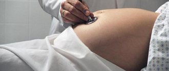
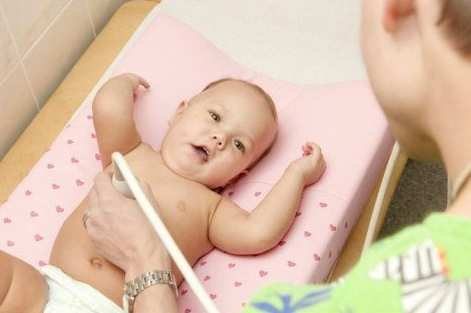
With the advent of ultrasound examinations, the number of diagnoses of diseases in the early stages of development has increased. This is very important in pediatrics, when it is difficult to determine pathology in newborns and young children based on clinical manifestations. Therefore, ultrasound for children is performed routinely at the age of up to 1 year, when the baby is growing rapidly. And any deviations in its development can affect the rest of his life.
Babies under 3 months should undergo an ultrasound of the abdomen, brain and joints. Diagnosis of the heart, kidneys or other organs is carried out if indicated.
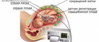
Ultrasound of the heart in children
Cardiac ultrasound is performed in newborns to exclude congenital heart defects in the presence of a genetic predisposition or indication. In other cases, ultrasound examination is prescribed for children after 12 years of age. Its goal is to identify those changes in the heart that are not detected by an ECG and are not accompanied by pain.
Ultrasound of the heart allows:
- Examine the lining of the heart, chambers and valve apparatus, the state of the myocardium - the muscles of the heart;
- Identify myocardial thickening;
- Identify congenital or acquired heart defects;
- Valve pathology;
- Ischemic disease;
- Aneurysm;
- Neoplasms, blood clots, chords and trabeculae, additional intracardiac formations.
Preparing for the cardiac ultrasound procedure
In order to accurately determine whether there is a pathology or not, you need to prepare. Of course, no special preparation is required here, but some rules will have to be followed before the examination:
- The day before the ultrasound, you do not need to drink alcoholic beverages, strong tea or coffee.
- It is not recommended to smoke on the day of the procedure, and if you cannot completely give up nicotine, then you should not smoke at least a couple of hours before the examination.
- If you regularly take any medications, be sure to notify your cardiologist in advance, who may ask you to refrain from taking them on the day of the examination.
- 10 minutes before the start of the ultrasound, you should sit down, rest and try to relax as much as possible.
- In the office where the procedure will be carried out, you should take with you a sheet, which you will lay on the couch, and a towel, with which you will have to wipe off the remaining gel.
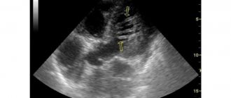
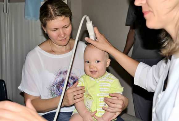
Ultrasound of the brain in children
Neurosonography is an ultrasound examination that is possible in a limited period of time. It is performed on a newborn as planned or when indicated until the large fontanel on the baby’s head has closed, which subsequently prevents the penetration of ultrasound. As a rule, this occurs before 1 year of age, but in individual cases the fontanelles can close in the first 6 months of life.
Indications for ultrasound of the brain:
- Complicated pregnancy;
- Difficult labor, birth asphyxia;
- Premature birth;
- Intracranial hemorrhage during childbirth;
- Injuries of the skull, brain;
- Anomalies in development, including the brain;
- Persistent neurological symptoms;
- Increased intracranial pressure;
- Unusual structure of the skull or face;
- Intrauterine infection.
Neurosonography allows you to study the structure of the child’s brain, determine structural changes, hemorrhages, ischemic lesions, and cysts. Ultrasound diagnoses the accumulation of cerebrospinal fluid in the ventricles of the brain in the early stages. Identify pathologies of the central nervous system. And in combination with Doppler ultrasound, it is possible to assess blood flow in the arteries of the brain.
Perform an ultrasound on the child
Interpretation of ultrasound of the heart of newborns
The interpretation of ultrasound data is carried out by an ultrasound specialist. The data obtained are compared with a table of age norms and a conclusion is drawn, which indicates the structure and size of the heart, and the volume of blood flow through the valves. Assess the functionality of the heart muscle.
Normal EchoCG indicators for a newborn baby:
- left ventricular ejection fraction 65 to 76 percent;
- left ventricular wall thickness up to 4.6 mm;
- the septum between the ventricles is from 4 to 9 mm;
- the wall thickness of the right ventricle is up to 3.4 mm.
The indicators may vary and be slightly distorted, it all depends on which machine the ultrasound is performed on. The most accurate results are provided by modern equipment, which can be used to obtain 4D and 3D images.
Ultrasound of the kidneys in children
Ultrasound of the kidneys is prescribed for newborns up to 3 months in order to exclude developmental abnormalities and for children, if indicated. Children whose parents suffer from chronic kidney disease are at risk for kidney and urinary tract pathologies. You should also be more attentive to girls who are physiologically more susceptible to urinary tract and kidney infections.
Indications for ultrasound of the kidneys:
- Genetic predisposition to kidney pathologies;
- Suspicion of congenital defects;
- Unsatisfactory urine tests;
- Abdominal and back injuries;
- Pain in the kidney area when urinating.
During an ultrasound of the kidneys, the shape, size and structure of the paired organ, the location of the kidneys and their functional state are assessed.
Ultrasound of the abdominal cavity in children
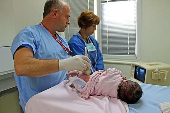
For the first time, ultrasound of the abdominal organs is performed on infants at 1 month. The purpose of the study is to assess the size of the abdominal cavity, the structure and relative position of organs, to identify anomalies in their development, foci of inflammatory processes and additional formations.
Ultrasound of the abdominal organs examines:
- Liver;
- Gallbladder;
- Bile ducts;
- Pancreas;
- Spleen;
- Intestines;
- Thymus gland;
- Retroperitoneal space – lymph nodes, great vessels.
ultrasound of a month old baby
Good afternoon Help me please. We haven't been able to conceive a child for over a year. Ultrasound of the pelvic organs: Uterus in anteflexio. The size of the uterus is 30*26*32mm. Cervix b/o. 10/11/13 7 d/c Right ovary 34*19*26mm follicle 9mm, w/body - Lev. ovary 35*16*27mm follicle 8mm, g/body - Endometrium - 3mm 10/16/13. 12d/c Right ovary follicle 8mm, left 8mm, endometrium - 6.5mm. No free fluid was detected. Conclusion: Echo signs of moderate uterine hypoplasia and signs of insufficient follicle maturation. Tests: 1. Progesterone 0.99 (normal folic phase 0.2-1.5 ng/ml, ovulatory phase 0.8-3 ng/ml) 2. Testosterone 0.50 (normal 0.06-0.82 ng /ml) 3. Prolactin 4028.3 (normal 60-600 mIU/l) 4. Estradiol 0.2 (normal 0.14-0.7 nmol/l) MRI excluded pituitary adenoma.
Ultrasound of the thyroid gland: In the right lobe 3 cm3, In the left lobe 3 cm3
TSH tests - 12.7 (normal 0.3-4 µIU/ml) T4free. - 7.9 (normal 9 - 22.2 pmol/l) T3free. - 1 (norm 1.2-4.2 pg/ml) A/t to TG - 10.7 (norm up to 100IU/ml) A/t to TPO - 9.7 (norm up to 30IU/ml) Conclusion: Hypoplasia thyroid gland 1 tbsp. Hypothyroidism.
Treatment was prescribed by an endocrinologist: 1) Eutirox 75 1 t/day (my weight is 55 kg) 2) Iodomarin 100 1 t/day 3) Folic acid 2 tablets. morning, 2 t/evening 4) Selenium 100 1t/day 2 months 5) Bromocriptine - 1t/morning, 1t/evening 6) Vitamin E 400 1t/day 1 month.
Please tell me if the treatment is prescribed correctly. Will it be possible to conceive, carry and give birth to a healthy child with such indicators? And how long does it take to be treated?
Thank you in advance
Have a nice day! Finally, these 2 months have passed. I passed the tests after two months of treatment. I took: 1) Eutirox 75 1 t/day (my weight is 55 kg) - 1 month, from the second month I started taking Eutirox at the rate of 1.6 * kg of weight, i.e. 1 table 75+ 1/4 tablets = 93.75
2) Iodomarin 100 1t/day 3) Folic acid 2 tablets. morning, 2 t/evening 4) Selenium 100 1t/day for 2 months 5) Bromocriptine - 1t/morning, 1t/evening - 1 month, from the second month, I started taking 1/2 tablet in the morning and evening, and then discontinued. 6) Vitamin E 400 1t/day for 1 month.
Here are the results of my tests after 2 months of treatment:
TSH - 0.1 µIU/ml, normal (0.3-4) T4free. — 18.2 pmol/l, normal (9-22.2) Prolactin — 1064.1 mMol/l, normal (60-600)
Please tell me what to do next in treatment? Is it necessary to reduce the dosage of eutirox? did it fall below the minimum value??? Prolactin is still too high. Is it possible to plan a baby or is it not yet possible with such prolactin levels? Should I continue to take folic acid and iodomarin?
Ultrasound of joints in children
In newborns, ultrasound of the joints should be performed routinely at 1 month. In the future, the study is carried out when indicated by a referral from an orthopedic surgeon after a scheduled examination at 6, 9, 12 months.
Indications for ultrasound of joints for children in the first year of life:
- Premature babies, complicated childbirth;
- Breech presentation;
- Limited lateral hip abduction;
- Different lengths of the lower limbs;
- Asymmetry of skin folds;
- Hypertonicity of the muscles of the lower extremities.
Ultrasound of joints in children allows timely detection of pathology of the hip joints, dislocations, subluxations, injuries during childbirth, developmental delays and dysplasia - underdevelopment of the joints. An accurate diagnosis in the early stages allows for timely treatment of disorders even before their clinical manifestations. In most cases, this approach allows you to completely cope with the pathology.
Ultrasound allows you to visualize not only bone tissue and joint spaces, but also tendons, ligaments, cartilage, joint capsule and synovial membrane. Medical recommends ultrasound examination using modern equipment with high resolution.
Types of ultrasound examination
There are several types of ultrasound that are performed on children aged 1 month.
Hip examination
This study is carried out after the child is 1 month old, but not later than he reaches 2 months. At this time, disorders are often discovered that were not noticed immediately after birth.
This procedure allows you to diagnose dysplasia of the hip joint, as well as conditions of its dislocation, which appeared during the development of the baby in the womb and during childbirth.
The sooner the pathology is diagnosed, the easier it is to correct the disorder, which helps to fully restore the musculoskeletal system of children. If treatment was started in the first month of a child’s life, it can be completely cured by six months. If treatment procedures began to be performed after 3 months, the pathology may subside only after 12 months, and surgery is often necessary.
If parents have not provided their child with the necessary treatment, over time he may develop movement disorders, and sometimes develop early arthrosis.
Brain research
This is a routine examination that is required for all children aged 1 month. The purpose of this study is to diagnose pathologies that appeared after fontanel closure. In infants, the cranial bones are not yet fully formed; they do not touch each other tightly and form fontanelles that can easily transmit ultrasonic waves. Ultrasound promotes active research into the structure of the child’s brain, detects congenital pathologies, as well as diseases that were acquired during intrauterine development or during labor.
This study allows you to diagnose:
- increase in intracranial pressure;
- the appearance of cysts;
- the presence of congenital malformations;
- formation of hydrocephalus;
- the appearance of intracranial hemorrhages.
Abdominal examination
To diagnose congenital pathologies, ultrasound is routinely prescribed to all babies. The procedure analyzes the proportionality to the age characteristics of the pancreas, liver, spleen, duodenum, and gall bladder.
Congenital diseases and the appearance of neoplasms in the abdominal cavity in children are also diagnosed.
Kidney examination
The need for a kidney examination is determined by a physician. This procedure is sometimes carried out until the baby is one month old. This is due to the fact that infants sometimes develop hydronephrosis of the kidney, which requires immediate surgical intervention. Also, the baby may additionally be prescribed screening of the abdominal cavity and stomach if he suffers from frequent regurgitation or lacks body weight.
Heart study
Such diagnostics should be carried out for every child until he reaches the twelfth month of life.

This type of ultrasound provides complete information about the health of the heart valves and blood vessels and developing cardiac pathologies.
Spinal region examination
If a birth injury is suspected, an ultrasound of the cervical spine is prescribed. This type of spine diagnosis is performed on babies every month. Also, examination of the cervical spine is necessary for muscular torticollis.
Cervical spine screening is a completely safe process that does not involve radiation exposure.
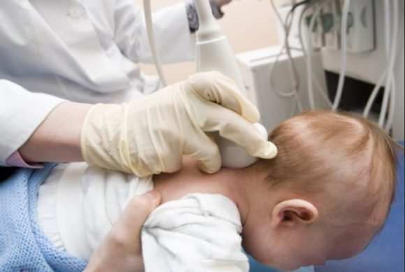
This procedure can be carried out several times to make an accurate diagnosis without harm to the baby.
Typically, the cervical spine screening procedure should be done:
- to identify birth injuries of the upper spine in children in the first month of life;
- in the presence of decreased muscle tone in the upper extremities;
- if during childbirth the fetus was entwined with the umbilical cord;
- if you have regular headaches;
- when unpleasant pain occurs in the cervical spine with light pressure;
- if you suspect early osteochondrosis;
- in the presence of domestic injuries in the cervical spine.